Table of Contents

Innate immune response and Th1 inflammation

Introduction

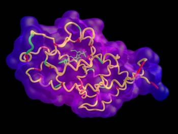
The innate immune responseThe body's first line of defense against intracellular and other pathogens. According to the Marshall Pathogenesis the innate immune system becomes disabled as patients develop chronic disease. is the body's first line of defense against and a non-specific way for responding to bacterial pathogens.1) Located in the nucleus of a variety of cells, the Vitamin D nuclear receptorA nuclear receptor located throughout the body that plays a key role in the innate immune response. (VDRThe Vitamin D Receptor. A nuclear receptor located throughout the body that plays a key role in the innate immune response.) plays a crucial, often under-appreciated, role in the innate immune response. The vitamin D receptor and T cell function

When functioning properly, the VDR transcribes between hundreds2) and thousands of genes3) including those for the proteins known as the antimicrobial peptidesBody’s naturally produced broad-spectrum antibacterials which target pathogens.. Antimicrobial peptides are “the body's natural antibiotics,” crucial for both prevention and clearance of infection.4) The VDR also expresses the TLR2A receptor which is expressed on the surface of certain cells and recognizes native or foreign substances and passes on appropriate signals to the cell and/or the nervous system. receptor, which is expressed on the surface of certain cells and recognizes foreign substances.

The body controls activity of the VDR through regulation of the vitamin D metabolites. 25-hydroxyvitamin D (25-D) antagonizes or inactivates the Receptor while 1,25-dihydroxyvitamin DPrimary biologically active vitamin D hormone. Activates the vitamin D nuclear receptor. Produced by hydroxylation of 25-D. Also known as 1,25-dihydroxycholecalciferol, 1,25-hydroxyvitamin D and calcitirol. (1,25-DPrimary biologically active vitamin D hormone. Activates the vitamin D nuclear receptor. Produced by hydroxylation of 25-D. Also known as 1,25-dihydroxycholecalciferol, 1,25-hydroxyvitamin D and calcitirol.) agonizes or activates the Receptor.

Greater than 36 types of tissue have been identified as having a Vitamin D Receptor.5)

Another component of the innate immune response is the release of inflammatory cytokinesAny of various protein molecules secreted by cells of the immune system that serve to regulate the immune system.. The result is what medicine calls inflammationThe complex biological response of vascular tissues to harmful stimuli such as pathogens or damaged cells. It is a protective attempt by the organism to remove the injurious stimuli as well as initiate the healing process for the tissue., which generally leads to an increase in symptoms.

Adenosine: an endogenous regulator of innate immunityThe body's first line of defense against intracellular and other pathogens. According to the Marshall Pathogenesis the innate immune system becomes disabled as patients develop chronic disease.. 6)

Before the Human MicrobiomeThe bacterial community in the human body. Many species in the microbiota contribute to the development of chronic disease. Project, scientists couldn't link bacteria to inflammatory diseases. But with the advent of DNA sequencing technology, scientists have detected many of the bacteria capable of generating an inflammatory response. All diseases of unknown etiology are inflammatory diseases.

Critical illness is a complex life-threatening disease characterized by profound endocrine and metabolic alterations and by a dysregulated immune response, together contributing to the susceptibility for nosocomial infections and sepsis

Hyperglycaemia, as part of the endocrine-metabolic responses to stress, is present in virtually all critically ill patients and is associated with poor outcome.

Not using parenteral nutrition during the first week in intensive care units, and so accepting a large macronutrient deficit, also resulted in fewer secondary infections, less weakness and accelerated recovery.

The human body: how we fail how we heal

Research

Those with a high intellectual capacity (hyper brain) possess overexcitabilities in various domains that may predispose them to certain psychological disorders as well as physiological conditions involving elevated sensory, and altered immune and inflammatory responses (hyper body). Science Direct

Modern vaccinations, fear of germs and obsession with hygiene are depriving the immune system of the information input upon which it is dependent. ….and may lead to increased incidences of allergies and autoimmune diseases. 7)

Country kids had more, and more-diverse, bacteria on their skin, with a particularly high abundance of Acinetobacter—a genus of microbes in the Proteobacteria phylum that are commonly found on plants. The researchers further found that children with more Acinetobacter on their skin had more leukocytes in their bloodstream and that these cells were much more capable of producing the anti-inflammatory cytokineAny of various protein molecules secreted by cells of the immune system that serve to regulate the immune system. IL-10 compared with the leukocytes of urban kids. influence of soil on human health

Insulin gives an extra boost to the immune system 8)

Results indicate a relationship between muscular TLR4, p-AMPK and NF-κB content and insulin sensitivity. The study also highlights that in situations of insulin resistance, such as in diabetic subjects, metformin treatment may prevent attenuation of activation of the inflammatory pathway. 9)

Glucose homeostasis, nutrition and infections during critical illness 10)

Adenosine is an important molecule that exerts control on the immune system, by signaling through receptors lying on the surface of immune cells. 11)

An exciting area of recent investigation has arisen from the discovery that SCFA play a role in regulating the immune system and inflammatory response. Early work at the turn of this century had demonstrated the potential role of butyrate in immune regulation when it was shown that butyrate inhibits nuclear factor kappa β (NF-κΒ) activation in macrophages and also inhibits histone deacetylation (HDAc) in acute myeloid leukemia.

Whether SCFA act as a signal to induce tolerance to the host-associated microbiome or directly reduce inflammatory responses remains to be fully elucidated. SCFA do appear able to reduce the responsiveness of lamina propria macrophages to commensal bacteria, via nitric oxide, IL-6, and IL-12 independent of FFAR signaling, to induce tolerance.

Recent data also suggests butyrate suppresses TNF-α, IL-6, and myeloperoxidase activity by preventing NF-κΒ activation in Kupffer cells.12)

Nuclear receptors and ligands

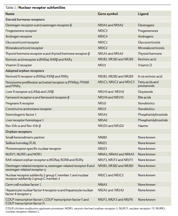
Nuclear receptorsIntracellular receptor proteins that bind to hydrophobic signal molecules (such as steroid and thyroid hormones) or intracellular metabolites and are thus activated to bind to specific DNA sequences which affect transcription. are a class of proteins found within the interior of cells that are responsible for sensing the presence of hormones and certain other molecules. A unique property of nuclear receptors which differentiate them from other classes of receptors is their ability to directly interact with and control the expression of genomic DNA. Some of the molecules (or ligands) which bind the nuclear receptorIntracellular receptor proteins that bind to hydrophobic signal molecules (such as steroid and thyroid hormones) or intracellular metabolites and are thus activated to bind to specific DNA sequences which affects transcription. activate (agonize) it and some inactivate (antagonize) it.

It is commonly accepted that most ligands, approximately 95% to 98%, inactivate the nuclear receptors. Since the nuclear receptors play a significant role in the immune response, this factor alone may explain why so many drugs and substances found in food and drink are immunosuppressive.

Because the expression of a large number of genes is regulated by nuclear receptors, ligands that activate these receptors can have profound effects on the organism. Many of these regulated genes are associated with various diseases which explains why the molecular targets of approximately 13% of FDA approved drugs are nuclear receptors.13)

Different cell types have different nuclear receptors. One of the nuclear receptors seen in immune cells is the Vitamin D Receptor (VDR). The VDR has two endogenous or “native” ligands, which are also the two main forms of vitamin D in the human body: 25-hydroxyvitamin D (25-D) and 1,25-dihydroxyvitamin D (1,25-D). Non-native or exogenous ligands can also inactivate or activate a nuclear receptor, depending on its molecular structure.

Ligands compete to dock at nuclear receptors. When is a given kind of ligand such as 25-D as opposed to 1,25-D more likely to bind to the VDR? It depends. 1,25-D tends to be much less common than 25-D – by a factor of 1,000 or more – so it binds to the receptor much more infrequently. A greater concentration of a given molecule can displace competing molecules off the nuclear receptor. Affinity occurs in logarithmic fashion, which is to say that it operates on the basis of a sliding scale. In short, an increase in 1,25-D and a decrease in 25-D can tilt the odds in favor of 1,25-D, and vice versa.

Affinity as well as the question of whether a ligand inactivates or activates a nuclear receptor can all be validated using in silicoExperiment technique performed on computer or via computer emulation. modeling. Although less precise, it is also possible to measure these properties in vitroA technique of performing a given procedure in a controlled environment outside of a living organism - usually a laboratory..

Activated by 1,25-D and inactivated by 25-D, the Vitamin D nuclear receptor (VDR) transcribes a number of genes crucial to the function of the innate immune response.

Role of Vitamin D Receptor in innate immunity

Vitamin D/VDR have multiple critical functions in regulating the response to intestinal homeostasis, tight junctions, pathogen invasion, commensal bacterial colonization, antimicrobe peptide secretion, and mucosal defense…. The involvement of Vitamin D/VDR in anti-inflammation and anti-infection represents a newly identified and highly significant activity for VDR.

Jun Sun 14)

When activated by 1,25-D, the Vitamin D Receptor (also called the calcitriol receptor) transcribes thousands of genes.15) It is commonly known that the VDR functions in regulating calcium metabolism.16) It is becoming increasingly clear, however, that the clinically accepted role of the Vitamin D metabolites, that of regulating calcium homeostasis, is just a small subset of the functions actually performed by these hormones.

Transcription of antimicrobial peptides

One of the VDR's key functions is the transcription of antimicrobial peptides.17) 18) See below.

Other antimicrobial activity of the VDR

Additionally, when the VDR is activated, TLR2 is expressed.19) TLR2 is a receptor, which is expressed on the surface of certain cells and recognizes native or foreign substances, and passes on appropriate signals to the cell and/or the nervous system.

When activated TLR2 allows the immune system to recognize gram-positive bacteria, including Staphylococcus aureus20) 21) Chlamydia pneumoniae22) and Mycoplasma pneumoniae.23) TLR2 also protects from intracellular infections such as Mycobacteria tuberculosis.24)

Antimicrobial peptides

The antimicrobial peptides (AMPs), of which there are hundreds, are families of proteins, which have been called “the body's natural antibiotics,” crucial for both prevention and clearance of infection. AMPs are broad-spectrum, responding to pathogens in a non-specific manner.25)

For example, consider cathelicidin Family of antimicrobial peptides found primarily in immune cells and transcribed by the Vitamin D Receptor., a protein transcribed by the VDR, which not unlike a Swiss Army knife, has many different functions. Because it can be differentially spliced, the cathelicidin protein itself can respond to a range of very different microbial challenges. In humans, the cathelicidin antimicrobial peptide gene encodes an inactive precursor protein (hCAP18) that is processed to release a 37amino-acid peptide (LL-37) from the C-terminus. LL-37 is susceptible to proteolitic processing by a variety of enzymes, generating many different cathelicidin-derived peptides, each of which has specific targets. For example, LL-37 is generated in response to Staphylococcus aureus, yet LL-37 represents 20% of the cathelicidin-derived peptides, with the smaller peptides being much more abundant and able to target even more diverse microbial forms.26)

AMPs have been documented to kill bacteria and disrupt their function through the following modes of action:

Also, AMPs aid in recovery from infection by:

In many cases, the exact mechanism by which antimicrobial peptides kill bacteria is unknown. In contrast to many conventional antibiotics including those used by the Marshall ProtocolA curative medical treatment for chronic inflammatory disease. Based on the Marshall Pathogenesis., AMPs appear to be bacteriocidal (a killer of bacteria) instead of bacteriostatic (an inhibitor of bacterial growth).

Two of the more significant families of AMPs are cathelicidin and the beta-defensins. Of these two families, cathelicidin is the most common.

Cathelicidin is a “fire alarm”, generating protective NLRP3-dependent airway epithelial cell inflammatory responses 27)

The full extent by which microbes interfere with AMP expression is the subject of a rapidly growing body of research.28) 29) 30)

Recent research into valuable proteins

Whey proteins and numerous growth factors that regulate insulin secretion, differentiation of intestine epithelium cells, and also tissue restoration, are priceless in stimulation the immune system.

Lactoferrin shows the most comprehensive pro-health properties: antioxidative, anticancer, immune stimulative and even chemopreventive. Also peptides and amino acids formed from casein and whey proteins possess immune stimulative activity. The most valuable proteins, i.e. lactoferrin, immune globulins, lactoperoxidase and lisozyme, together with bioactive peptides, are resistant to pepsin and trypsin activity. This is why they maintain their exceptional biological activity. 31)

Antimicrobial peptides target fungi and viruses

The antimicrobial peptides play a role in mitigating the virulence of the virome and other non-bacterial infectious agents. In addition to its antibacterial activity, alpha-defensin human neutrophil peptide-1 inhibits HIV and influenza virus entry into target cells.32) It diminishes HIV replication and can inactivate cytomegalovirus, herpes simplex virus, vesicular stomatitis virus and adenovirus.33) In addition to killing both gram positive and gram-negative bacteria, human beta-defensins HBD-1, HDB-2, and HBD-3 have also been shown to kill the opportunistic yeast species Candida albicans.34) Cathelicidin also possesses antiviral and antifungal activity.35) 36)

In other words, there is a reason why this group of proteins are named antimicrobial peptides rather than antibacterial peptides.

Unexpected antimicrobial peptides

There are now several examples of substances believed to cause disease, which have since been proven to be part of host defense.

Evolutionarily conserved

The TLR2/1 and cathelicidin-vitamin D pathway has long played a “powerful force” in protecting the body against infection. This is evidenced by the fact that the Alu short interspersed element (SINE), which transcribes the vitamin D receptor binding element (VDRE), has been evolutionarily conserved for 55-60 million years, but not prior.39) The differences in this pathway between humans/primates and other mammals call into question animal models that try to emulate the vitamin D system and indeed the immune system.

Energy consumption

It has become clear in recent years that pretty much any immune cell in our body undergoes, upon its activation, a metabolic shift resembling the Warburg effect originally described for cancer cells. Immune cells increase glucose consumption and produce a significant portion of ATP by glycolysis ending with lactate even under oxygenated conditions; increased glycolysis is required for the generation of intermediate metabolites associated with the activation of the immune cell.

Increased energy consumption by immune cells requires a metabolic adaptation of the whole organism. During trauma or infection, the organism vitally depends on the immune system, which is therefore privileged in energy/nutrient allocation. According to Rainer Straub [2], insulin resistance caused by pro-inflammatory cytokines is a physiological way of the immune system to usurp energy/nutrients during acute stress from the rest of the organism because immune cells themselves do not become insulin resistant. 40)

Inflammation

Another component of the innate immune response is inflammation, the universal initial response of the organism to any injurious agent.41) Inflammation is a systemic physiological process fundamental for survival.42) The identification of bacteria and other pathogens triggers the release of inflammatory cytokines. These cytokines include interferon-gammaAn inflammatory cytokine which causes extra mast cells to differentiate to monocytes and then to further differentiate into macrophages and dendritic cells. These phagocytes are the most active cells of the immune system and are charged with digesting bacterial pathogens., tumor necrosis factor-alphaA cytokine critical for effective immune surveillance and is required for proper proliferation and function of immune cells. (TNF-alphaA cytokine critical for effective immune surveillance and is required for proper proliferation and function of immune cells.), and Nuclear Factor-kappa BA protein that stimulates the release of inflammatory cytokines in response to infection (NF-kappaBA protein that stimulates the release of inflammatory cytokines in response to infection). Cytokines are regulatory proteins, such as the interleukins and lymphokines, that are released by cells of the immune system and act as intercellular mediators in the generation of an immune response. The result is what medicine calls inflammation, which generally leads to an increase in symptoms.

Th1/Th17 inflammation

One key type of inflammation is the Th1/Th17 (T-helper) inflammatory response. In the interests of concision, the Th1/Th17, on this site and others, the Th1/Th17 response is referred to as the Th1 response. This reaction occurs in response to intracellular pathogens, which according to the Marshall Pathogenesis, play a driving force in chronic disease.

All Th1 diseases are marked by an inflammatory response

Before the Human Microbiome Project, scientists couldn't consistently link bacteria to inflammatory diseases. But with the advent of DNA sequencing technology, scientists have detected many of the bacteria capable of generating an inflammatory response. All diseases of unknown etiology are inflammatory diseases.

An inflammatory immune response—one of the body’s primary means to protect against infection—defines multiple established infectious causes of chronic diseases, including some cancers. Inflammation also drives many chronic conditions that are still classified as (noninfectious) autoimmuneA condition or disease thought to arise from an overactive immune response of the body against substances and tissues normally present in the body or immune-mediated (e.g., systemic lupus erythematosus, rheumatoid arthritis, Crohn’s disease). Both [the innate and adaptive immune systems] play critical roles in the pathogenesis of these inflammatory syndromes. Therefore, inflammation is a clear potential link between infectious agents and chronic diseases.

Siobhán M. O'Connor et al. 43)

Th2 inflammation

According to the Marshall Pathogenesis, generally speaking, any activity of the Th2 cytokines in chronic disease is a result of the primary Th1-inducing pathogens.

Many palliative therapies interfere with inflammation

While inflammation is associated with disease, inflammation often serves an invaluable role as the immune system fights off chronic pathogens. Numerous medications artificially suppress inflammation including anti-TNF drugsDrugs which interfere with the body's production of TNF-alpha - a cytokine necessary for recovery from infection, interferon, corticosteroidsA first-line treatment for a number of diseases. Corticosteroids work by slowing the innate immune response. This provides some patients with temporary symptom palliation but exacerbates the disease over the long-term by allowing chronic pathogens to proliferate., antifungals, and anti-pyreutics. While interfering with the inflammatory response typically reduces immunopathologyA temporary increase in disease symptoms experienced by Marshall Protocol patients that results from the release of cytokines and endotoxins as disease-causing bacteria are killed. and makes a patient feel less symptomatic in the near term, doing so allows the bacteria which cause chronic disease to proliferate.

The release of cytokines appears to be essential for recovery after an infection. One study found that the cytokine TNF-alpha – which is blocked by anti-TNF drugs – is necessary for the proper expression of acquired specific resistance following infection with Mycobacterium tuberculosis.44) 45) 46) Another effect of the use of TNF blockers is to break or reduce the formation of granuloma, one of the body's mechanisms to control bacterial pathogens.47)

Commensal microbes

The host innate immune defense system is highly active in healthy tissue.48) Commensal bacteria can activate innate immune responses.49) 50)

Immune suppression

The surprising finding of science is that home and hospital environments can be less supportive of human health than the outdoors. exposure to environmental microbes helps protect against allergies and other inflammatory diseases 51) Exposure to environmental microbes helps protect against allergies and other inflammatory diseases

Environmental pollution, both visible and invisible, many palliative medications, and increasingly this century the cellular level radiation from wifi devices, are interfering with the capacity of the innate immune system to create the defensive molecules necessary to control intracellular infections

===== Notes and comments =====

  • Legacy content
    • e92:

Vitamin D, Vitamin D Receptor, and Macroautophagy in Inflammation and Infection

https://tiny.cc/9xtyw

Cool paper, have a look at the references

This paper helps my understanding of innate immune action where I had questions. If it is correct and my understanding of the paper is good, then it looks like general cells use a VDR mediated house cleaning mechanism, autophagy, to deal with intracellular pathogens. This has significant implication to healing if it is correct and if it is turned on by olmesartanMedication taken regularly by patients on the Marshall Protocol for its ability to activate the Vitamin D Receptor. Also known by the trade name Benicar. . I think it means that generally, any infected nucleated cell could clear its intracellular pathogens if the intracellular infection has not blocked VDR creation. It would mean healing could occur without apoptosis of infected cells. That is what it seems to me that the article is implying.

On other points, the paper uses D3 and 1,25D3 interchangeably (because it is the active ligand form of D3), which makes me nervous as researchers usually use D3 and 25D3 interchangeably. I am also not confidant that the papers author knows that murine models are misleading when discussing human VDR.

In general, I am with Chris and would like some help with understanding this paper and its drawbacks.

Makes sense, we've bantered about autophagy in prior threads. The VDR may not be expressed all tissues and red blood cells are not nucleated.

This underscores the need to have s healthy immune syatem.

https://www.reuters.com/article/2011/04/22/us-biogen-tysabri-idUSTRE73L2WN20110422

“Limited data is available beyond four years”

..Trevor..

Cell Microbiol. 2010 Nov;12(11):1648-65. doi: 10.1111/j.1462-5822.2010.01497.x. Epub 2010 Jul 20. Mycobacterial lipoprotein activates autophagy via TLR2/1/CD14 and a functional vitamin D receptor signalling. Shin DM, Yuk JM, Lee HM, Lee SH, Son JW, Harding CV, Kim JM, Modlin RL, Jo EK. Department of Microbiology, Chungnam National University School of Medicine, Daejeon, Korea. Abstract In human monocytes, Toll-like receptor (TLR) 2/1 activation leads to vitamin D3-dependent antimycobacterial activities, but the molecular mechanisms by which TLR2/1 stimulation induces antimicrobial activities against mycobacteria remain unclear. Here we show that TLR2/1/CD14 stimulation by mycobacterial lipoprotein LpqH can robustly activate antibacterial autophagy through vitamin D receptor signalling activation and cathelicidin induction. We found that CCAAT/enhancer-binding protein (C/EBP)-β-dependent induction of 25-hydroxycholecalciferol-1α-hydroxylase (Cyp27b1) hydroxylase was critical for LpqH-induced cathelicidin expression and autophagy. In addition, increases in intracellular calcium following AMP-activated protein kinase (AMPK) activation played a crucial role in LpqH-induced autophagy. Moreover, AMPK-dependent p38 mitogen-activated protein kinase (MAPK) activation was required for LpqH-induced Cyp27b1 expression and autophagy activation. Collectively, these data suggest that TLR2/1/CD14-Ca(2+) -AMPK-p38 MAPK pathways contribute to C/EBP-β-dependent expression of Cyp27b1 and cathelicidin, which played an essential role in LpqH-induced autophagy. Furthermore, these results establish a previously uncharacterized signalling pathway of antimycobacterial host defence through a functional link of TLR2/1/CD14-dependent sensing to the induction of autophagy. © 2010 Blackwell Publishing Ltd. PMID: 20560977

Mycobacterial lipoprotein activates autophagy via TLR2/1/CD14 and a functional vitamin D receptor signalling https://www.ncbi.nlm.nih.gov/pmc/articles/PMC2970753/?tool=pubmed

“our findings clearly demonstrate that TLR2/1 signalling regulates antibacterial autophagy pathway through functional vitamin D3 receptor activation and cathelicidin expression in human primary monocytes.”

Nuclear hormone receptors as therapeutic targets.Levi M, Wang X, Choudhury D Contrib Nephrol 2011; 170() :209-16 Download citation Affiliation Division of Renal Diseases and Hypertension, Department of Medicine, University of Colorado Denver, Aurora, Colo., USA. Abstract In spite of excellent glucose and blood pressure control, including administration of angiotensin-converting enzyme inhibitors and/or angiotensin II receptor blockers, diabetic nephropathy (DN) still develops and progresses. The development of additional protective therapeutic interventions is, therefore, a major priority. Nuclear hormone receptors regulate carbohydrate metabolism, lipid metabolism, the immune response, inflammation and development of fibrosis. The increasing prevalence of DN has led to intense investigation of the role that nuclear hormone receptors may have in slowing or preventing the progression of renal disease. Several nuclear receptor-activating ligands (agonists) have been shown to have a renal protective effect in the context of DN. This review will discuss the evidence regarding the beneficial effects of the activation of the vitamin D receptor (VDR) and the farnesoid X receptor (FXR) in preventing the progression of DN, and will describe how the discovery and development of compounds that modulate the activity of VDR and FXR may provide potential additional therapeutic approaches in the management of DN.

Also include in “Mechanisms by which…”

Injury enhances TLR2 function and antimicrobial peptide expression through a vitamin D–dependent mechanism

https://www.ncbi.nlm.nih.gov/pmc/articles/PMC1784003/?tool=pubmed

An essential element of the innate immune response to injury is the capacity to recognize microbial invasion and stimulate production of antimicrobial peptides. We investigated how this process is controlled in the epidermis. Keratinocytes surrounding a wound increased expression of the genes coding for the microbial pattern recognition receptors CD14 and TLR2, complementing an increase in cathelicidin antimicrobial peptide expression. These genes were induced by 1,25(OH)2 vitamin D3 (1,25D3; its active form), suggesting a role for vitamin D3 in this process. How 1,25D3 could participate in the injury response was explained by findings that the levels of CYP27B1, which converts 25OH vitamin D3 (25D3) to active 1,25D3, were increased in wounds and induced in keratinocytes in response to TGF-β1. Blocking the vitamin D receptor, inhibiting CYP27B1, or limiting 25D3 availability prevented TGF-β1 from inducing cathelicidin, CD14, or TLR2 in human keratinocytes, while CYP27B1-deficient mice failed to increase CD14 expression following wounding. The functional consequence of these observations was confirmed by demonstrating that 1,25D3 enabled keratinocytes to recognize microbial components through TLR2 and respond by cathelicidin production. Thus, we demonstrate what we believe to be a previously unexpected role for vitamin D3 in innate immunity, enabling keratinocytes to recognize and respond to microbes and to protect wounds against infection.

“To further investigate the significance of increased CYP27B1 activation in skin injury, wounds from mice lacking the CYP27B1 enzyme were evaluated for induction of an innate immune response. CD14 is regulated by 1,25D3 (29), and in mouse wounds, CD14 was induced in wild-type animals but not in those lacking CYP27B1 (Figure (Figure5A).5A). In contrast, murine Camp, which is not regulated by 1,25D3 (7), was increased in wounds in both wild-type and CYB27B1-deficient animals”

In VivoA type of scientific study that analyzes an organism in its natural living environment. Activation of the Intracrine Vitamin D Pathway in Innate Immune Cells and Mammary Tissue during a Bacterial Infection. https://www.ncbi.nlm.nih.gov/pubmed/21124742

Numerous in vitro studies have shown that toll-like receptor signaling induces 25-hydroxyvitamin D(3) 1α-hydroxylase (1α-OHase; CYP27B1) expression in macrophages from various species. 1α-OHase is the primary enzyme that converts 25-hydroxyvitamin D(3) to 1,25-dihydroxyvitamin D(3) (1,25(OH)(2)D(3)). Subsequently, synthesis of 1,25(OH)(2)D(3) by 1α-OHase in macrophages has been shown to modulate innate immune responses of macrophages. Despite the numerous in vitro studies that have shown 1α-OHase expression is induced in macrophages, however, evidence that 1α-OHase expression is induced by pathogens in vivo is limited. The objective of this study was to evaluate 1α-OHase gene expression in macrophages and mammary tissue during an in vivo bacterial infection with Streptococcus uberis. In tissue and secreted cells from the infected mammary glands, 1α-OHase gene expression was significantly increased compared to expression in tissue and cells from the healthy mammary tissue. Separation of the cells by FACS9 revealed that 1α-OHase was predominantly expressed in the CD14(+) cells isolated from the infected mammary tissue. The 24-hydroxylase gene, a gene that is highly upregulated by 1,25(OH)(2)D(3), was significantly more expressed in tissue and cells from the infected mammary tissue than from the healthy uninfected mammary tissue thus indicating significant local 1,25(OH)(2)D(3) production at the infection site. In conclusion, this study provides the first in vivo evidence that 1α-OHase expression is upregulated in macrophages in response to bacterial infection and that 1α-OHase at the site of infection provides 1,25(OH)(2)D(3) for local regulation of vitamin D responsive genes.

I have attached a recent paper that discusses the previously unrecognized function of prions as anti-microbial agents, sharing certain similarities with archetypal anti-microbial peptides like LL-37. I seem to recall you mentioning that you had attended a presentation in Karolinska that discussed how “prion disease” could only exist against the backdrop of a TH1 state. Certainly, the observation that many prions are “infectious” can now be explained by your model. It would appear that prion production may simply be the body's reaction to infection, rather than the cause of “prion disease.”

From the paper:

“As mentioned above, prion protein is not only confined to the nervous system, but instead ubiquitously found in many other cells and tissues, and the physiological role for this protein are still enigmatic. In a previous study, it was reported that human keratinocytes express PrPc in vitro and during inflammatory skin disease [18]. Although that previous work was focusing on prion infectivity routes, our current findings on increased expression of PrP during wounding, together with the observation of its antimicrobial activity, clearly indicate that PrPs could have a previously undisclosed role in host defense. In this context, experiments with PrP deficient animals in infection models should be valuable in order to further delineate a possible role of PrP in innate defense.”52)

“Notably, PrP bound LPS similarly to human LL-37.”

Dan Rudderow

Some AMPs have activity against certain species of antibiotic-resistant bacteria.(11557478)

Hydrogen sulfide: a marker of inflammation: https://www.eurekalert.org/pub_releases/2010-08/tpco-reg082010.php https://onlinelibrary.wiley.com/doi/10.1111/j.1749-6632.2010.05556.x/full

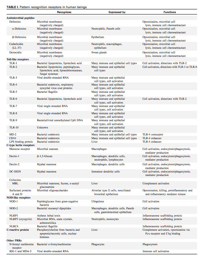
In a 2008 paper, Liu lists the different pattern recognition receptors in humans.53)

Clin Biochem. 2010 Jul 23. [Epub ahead of print] Correlation of a multi-cytokine panel with clinical disease activity in patients with rheumatoid arthritis.

Milman N, Karsh J, Booth RA.

Faculty of Medicine, University of Ottawa, 451 Smyth Rd., Ottawa ON, Canada K1H 8M5; Department of Medicine, Division of Rheumatology, 1967 Riverside Dr. Ottawa ON, Canada K1H 7W9. Abstract

OBJECTIVE: Explore the potential use of a cytokine panel as biochemical markers of disease activity in rheumatoid arthritis (RA) patients. DESIGN AND METHODS: 57 adult RA patients were assessed using five validated clinical disease activity tools: Health Assessment Questionnaire (HAQ), standard 28-joint Disease Activity Score (DAS28), DAS28 using C-reactive protein (DAS28-CRP), Clinical Disease Activity Index (CDAI), and Simple Disease Activity Index (SDAI). Plasma cytokine levels (IL-2, IL-4, IL-6, IL-8, IL-10, VEGF, IFN-gamma, TNF-alpha, IL1alpha, IL1beta, MCP1, and EGF) were measured in 47 of the 57 patients and correlated with clinical indicators. RESULTS: We found significant correlations between plasma levels of IL-6 and all clinical measures of disease activity; Spearman coefficients (p values) were: HAQ: 0.347(0.017); DAS28: 0.409(0.005); DAS-CRP: 0.378(0.011); CDAI: 0.312(0.033); SDAI: 0.310(0.039); ESR: 0.448(0.002); and CRP: 0.513(0.001). IFN-gamma also correlated with DAS-CRP: 0.309(0.039) and SDAI: 0.301(0.044). Furthermore, the levels of IL-6 and IFN-gamma increased significantly with worsening disease, as defined by the European League Against Rheumatism (EULAR) classification of disease activity. CONCLUSION: A significant correlation between plasma levels of IL-6 and clinical disease activity in patients with RA suggests a future role of IL6 as a disease activity marker. Copyright © 2010. Published by Elsevier Inc.

PMID: 20655893

Plasma cytokine fluctuations over time in healthy controls and patients with fibromyalgia.

Togo F, Natelson BH, Adler GK, Ottenweller JE, Goldenberg DL, Struzik ZR, Yamamoto Y.

Pain & Fatigue Study Center, Department of Neurosciences, UMDNJ-New Jersey Medical School, 1618 ADMC, 30 Bergen Street, Newark, NJ 07103, USA.

We examined the pattern of cytokine secretion across the 24-hr day for women with widespread pain and tenderness having the diagnosis of fibromyalgia (FM) and matched healthy controls. Subjects were given time to habituate to being in a clinical research laboratory environment and then were sampled for cytokines without their being disturbed for a 24-hr period including an 8-hr sleep period. Cytokine levels were uniformly low but characterized by bursts of secretion. Bursting occurred either in singlets or in doublets with a range from 88 to 131 mins between doublet bursts. There was an element of synchronization of these bursts with most occurring at the beginning of sampling. FM patients showed a shift to increased IL-10 in the nighttime compared to controls. The relation between this anti-inflammatory cytokine to the pro-inflammatory cytokines studied also differed between groups: FM patients showed an increased ratio of IL-10 burst amplitude to that of pro-inflammatory cytokines IL-1beta, IL-8, and TNF-alpha. We interpret this to indicate a skew away from the normal balance favoring pro-inflammatory cytokines in controls toward one favoring an anti-inflammatory response in FM. These changes toward anti-inflammatory predominance in FM may explain their common complaint of disturbed sleep because these cytokines are known to disrupt sleep.

PMID: 19064941 [PubMed - indexed for MEDLINE]

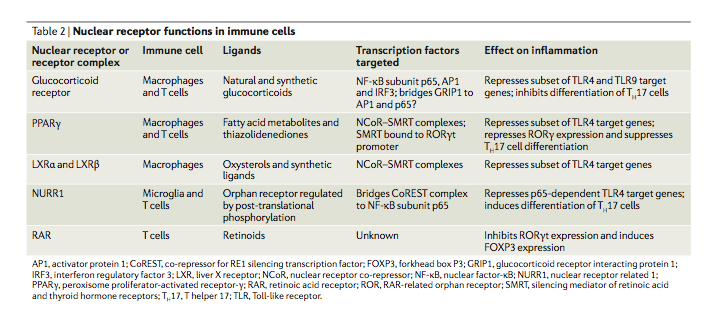
Nuclear receptor transrepression pathways that regulate inflammation in macrophages and T cells54)

  • Leukocytes (immune cells)
    • Adaptive Immune
    • Innate Immune
      • Mast Cells – resides in the connective tissue and in the mucous membranes, and are intimately associated with defense against pathogens, wound healing, but are also often associated with allergy and anaphylaxis.
      • Phagocytes – immune cells that engulf, i.e. phagocytose, pathogens or particles
        • Macrophages – large phagocytic leukocytes, which are able to move outside of the vascular system by moving across the cell membrane of capillary vessels and entering the areas between cells in pursuit of invading pathogens.
        • Neutrophils – contain granules in their cytoplasm; neutrophil granules contain a variety of toxic substances that kill or inhibit growth of bacteria and fungi
        • Dendritic cells – present in tissues that are in contact with the external environment, mainly the skin (where they are often called Langerhans cells), and the inner mucosal lining of the nose, lungs, stomach and intestines. They are named for their resemblance to neuronal dendrites, but dendritic cells are not connected to the nervous system. Dendritic cells are very important in the process of antigen presentation, and serve as a link between the innate and adaptive immune systems.
      • Basophils and eosinophils – When activated by a pathogen encounter, basophils releasing histamine are important in defense against parasites, and play a role in allergic reactions (such as asthma). Upon activation, eosinophils secrete a range of highly toxic proteins and free radicals that are highly effective in killing bacteria and parasites, but are also responsible for tissue damage occurring during allergic reactions
      • Natural killer cells – destroy compromised host cells, such as tumor cells or virus-infected cells, recognizing such cells by a condition known as “missing self.” This term describes cells with low levels of a cell-surface marker called MHC I (major histocompatibility complex) - a situation that can arise in viral infections of host cells. They were named “natural killer” because of the initial notion that they do not require activation in order to kill cells that are “missing self.”
      • gamma/delta T cells
      • monocytes – several roles: (1) replenish resident macrophages and dendritic cells under normal states, and (2) in response to inflammation signals, monocytes can move quickly (approx. 8-12 hours) to sites of infection in the tissues and divide/differentiate into macrophages and dendritic cells to elicit an immune response

===== References =====

1)
Rasmussen SB, Reinert LS, Paludan SR. Innate recognition of intracellular pathogens: detection and activation of the first line of defense. APMIS. 2009 May;117(5-6):323-37. doi: 10.1111/j.1600-0463.2009.02456.x.
[PMID: 19400860] [DOI: 10.1111/j.1600-0463.2009.02456.x]
2)
Ramagopalan SV, Heger A, Berlanga AJ, Maugeri NJ, Lincoln MR, Burrell A, Handunnetthi L, Handel AE, Disanto G, Orton S, Watson CT, Morahan JM, Giovannoni G, Ponting CP, Ebers GC, Knight JC. A ChIP-seq defined genome-wide map of vitamin D receptor binding: associations with disease and evolution. Genome Res. 2010 Oct;20(10):1352-60. doi: 10.1101/gr.107920.110. Epub 2010 Aug 24.
[PMID: 20736230] [PMCID: 2945184] [DOI: 10.1101/gr.107920.110]
3) , 15)
Wang T, Tavera-Mendoza LE, Laperriere D, Libby E, MacLeod NB, Nagai Y, Bourdeau V, Konstorum A, Lallemant B, Zhang R, Mader S, White JH. Large-scale in silico and microarray-based identification of direct 1,25-dihydroxyvitamin D3 target genes. Mol Endocrinol. 2005 Nov;19(11):2685-95. doi: 10.1210/me.2005-0106. Epub 2005 Jul 7.
[PMID: 16002434] [DOI: 10.1210/me.2005-0106]
4) , 25)
Zasloff M. Antimicrobial peptides of multicellular organisms. Nature. 2002 Jan 24;415(6870):389-95. doi: 10.1038/415389a.
[PMID: 11807545] [DOI: 10.1038/415389a]
5)
Norman AW. From vitamin D to hormone D: fundamentals of the vitamin D endocrine system essential for good health. Am J Clin Nutr. 2008 Aug;88(2):491S-499S. doi: 10.1093/ajcn/88.2.491S.
[PMID: 18689389] [DOI: 10.1093/ajcn/88.2.491S]
6)
Haskó G, Cronstein BN. Adenosine: an endogenous regulator of innate immunity. Trends Immunol. 2004 Jan;25(1):33-9. doi: 10.1016/j.it.2003.11.003.
[PMID: 14698282] [DOI: 10.1016/j.it.2003.11.003]
7) , 51)
Rook GA, Stanford JL. Give us this day our daily germs. Immunol Today. 1998 Mar;19(3):113-6. doi: 10.1016/s0167-5699(97)01204-8.
[PMID: 9540269] [DOI: 10.1016/s0167-5699(97)01204-8]
8)
Tsai S, Clemente-Casares X, Zhou AC, Lei H, Ahn JJ, Chan YT, Choi O, Luck H, Woo M, Dunn SE, Engleman EG, Watts TH, Winer S, Winer DA. Insulin Receptor-Mediated Stimulation Boosts T Cell Immunity during Inflammation and Infection. Cell Metab. 2018 Dec 4;28(6):922-934.e4. doi: 10.1016/j.cmet.2018.08.003. Epub 2018 Aug 30.
[PMID: 30174303] [DOI: 10.1016/j.cmet.2018.08.003]
9)
Peixoto LG, Teixeira RR, Vilela DD, Barbosa LN, Caixeta DC, Deconte SR, de Assis de Araújo F, Sabino-Silva R, Espindola FS. Metformin attenuates the TLR4 inflammatory pathway in skeletal muscle of diabetic rats. Acta Diabetol. 2017 Oct;54(10):943-951. doi: 10.1007/s00592-017-1027-5. Epub 2017 Aug 9.
[PMID: 28791487] [DOI: 10.1007/s00592-017-1027-5]
10)
Ingels C, Vanhorebeek I, Van den Berghe G. Glucose homeostasis, nutrition and infections during critical illness. Clin Microbiol Infect. 2018 Jan;24(1):10-15. doi: 10.1016/j.cmi.2016.12.033. Epub 2017 Jan 7.
[PMID: 28082192] [DOI: 10.1016/j.cmi.2016.12.033]
11)
de Oliveira Bravo M, Carvalho JL, Saldanha-Araujo F. Adenosine production: a common path for mesenchymal stem-cell and regulatory T-cell-mediated immunosuppression. Purinergic Signal. 2016 Dec;12(4):595-609. doi: 10.1007/s11302-016-9529-0. Epub 2016 Aug 25.
[PMID: 27557887] [PMCID: 5124006] [DOI: 10.1007/s11302-016-9529-0]
12)
Morrison DJ, Preston T. Formation of short chain fatty acids by the gut microbiota and their impact on human metabolism. Gut Microbes. 2016 May 3;7(3):189-200. doi: 10.1080/19490976.2015.1134082. Epub 2016 Mar 10.
[PMID: 26963409] [PMCID: 4939913] [DOI: 10.1080/19490976.2015.1134082]
13)
Overington JP, Al-Lazikani B, Hopkins AL. How many drug targets are there?. Nat Rev Drug Discov. 2006 Dec;5(12):993-6. doi: 10.1038/nrd2199.
[PMID: 17139284] [DOI: 10.1038/nrd2199]
14)
Sun J. Vitamin D and mucosal immune function. Curr Opin Gastroenterol. 2010 Nov;26(6):591-5. doi: 10.1097/MOG.0b013e32833d4b9f.
[PMID: 20639756] [PMCID: 2955835] [DOI: 10.1097/MOG.0b013e32833d4b9f]
16)
Li YC, Bolt MJ, Cao LP, Sitrin MD. Effects of vitamin D receptor inactivation on the expression of calbindins and calcium metabolism. Am J Physiol Endocrinol Metab. 2001 Sep;281(3):E558-64. doi: 10.1152/ajpendo.2001.281.3.E558.
[PMID: 11500311] [DOI: 10.1152/ajpendo.2001.281.3.E558]
17)
Wang T, Nestel FP, Bourdeau V, Nagai Y, Wang Q, Liao J, Tavera-Mendoza L, Lin R, Hanrahan JW, Mader S, White JH. Cutting edge: 1,25-dihydroxyvitamin D3 is a direct inducer of antimicrobial peptide gene expression. J Immunol. 2004 Sep 1;173(5):2909-12. doi: 10.4049/jimmunol.173.5.2909.
[PMID: 15322146] [DOI: 10.4049/jimmunol.173.5.2909]
18)
Gombart AF, Borregaard N, Koeffler HP. Human cathelicidin antimicrobial peptide (CAMP) gene is a direct target of the vitamin D receptor and is strongly up-regulated in myeloid cells by 1,25-dihydroxyvitamin D3. FASEB J. 2005 Jul;19(9):1067-77. doi: 10.1096/fj.04-3284com.
[PMID: 15985530] [DOI: 10.1096/fj.04-3284com]
19)
Schauber J, Dorschner RA, Coda AB, Büchau AS, Liu PT, Kiken D, Helfrich YR, Kang S, Elalieh HZ, Steinmeyer A, Zügel U, Bikle DD, Modlin RL, Gallo RL. Injury enhances TLR2 function and antimicrobial peptide expression through a vitamin D-dependent mechanism. J Clin Invest. 2007 Mar;117(3):803-11. doi: 10.1172/JCI30142. Epub 2007 Feb 8.
[PMID: 17290304] [PMCID: 1784003] [DOI: 10.1172/JCI30142]
20)
Takeuchi O, Hoshino K, Akira S. Cutting edge: TLR2-deficient and MyD88-deficient mice are highly susceptible to Staphylococcus aureus infection. J Immunol. 2000 Nov 15;165(10):5392-6. doi: 10.4049/jimmunol.165.10.5392.
[PMID: 11067888] [DOI: 10.4049/jimmunol.165.10.5392]
21)
González-Zorn B, Senna JPM, Fiette L, Shorte S, Testard A, Chignard M, Courvalin P, Grillot-Courvalin C. Bacterial and host factors implicated in nasal carriage of methicillin-resistant Staphylococcus aureus in mice. Infect Immun. 2005 Mar;73(3):1847-51. doi: 10.1128/IAI.73.3.1847-1851.2005.
[PMID: 15731086] [PMCID: 1064969] [DOI: 10.1128/IAI.73.3.1847-1851.2005]
22)
Cao F, Castrillo A, Tontonoz P, Re F, Byrne GI. Chlamydia pneumoniae--induced macrophage foam cell formation is mediated by Toll-like receptor 2. Infect Immun. 2007 Feb;75(2):753-9. doi: 10.1128/IAI.01386-06. Epub 2006 Dec 4.
[PMID: 17145941] [PMCID: 1828523] [DOI: 10.1128/IAI.01386-06]
23)
Chu HW, Jeyaseelan S, Rino JG, Voelker DR, Wexler RB, Campbell K, Harbeck RJ, Martin RJ. TLR2 signaling is critical for Mycoplasma pneumoniae-induced airway mucin expression. J Immunol. 2005 May 1;174(9):5713-9. doi: 10.4049/jimmunol.174.9.5713.
[PMID: 15843573] [DOI: 10.4049/jimmunol.174.9.5713]
24)
Carlos D, Frantz FG, Souza-Júnior DA, Jamur MC, Oliver C, Ramos SG, Quesniaux VF, Ryffel B, Silva CL, Bozza MT, Faccioli LH. TLR2-dependent mast cell activation contributes to the control of Mycobacterium tuberculosis infection. Microbes Infect. 2009 Jul-Aug;11(8-9):770-8. doi: 10.1016/j.micinf.2009.04.025. Epub 2009 May 13.
[PMID: 19442756] [DOI: 10.1016/j.micinf.2009.04.025]
26) , 33)
Auvynet C, Rosenstein Y. Multifunctional host defense peptides: antimicrobial peptides, the small yet big players in innate and adaptive immunity. FEBS J. 2009 Nov;276(22):6497-508. doi: 10.1111/j.1742-4658.2009.07360.x. Epub 2009 Oct 9.
[PMID: 19817855] [DOI: 10.1111/j.1742-4658.2009.07360.x]
27)
McHugh BJ, Wang R, Li H, Beaumont PE, Kells R, Stevens H, Young L, Rossi AG, Gray RD, Dorin JR, Gwyer Findlay EL, Brough D, Davidson DJ. Cathelicidin is a "fire alarm", generating protective NLRP3-dependent airway epithelial cell inflammatory responses during infection with Pseudomonas aeruginosa. PLoS Pathog. 2019 Apr 12;15(4):e1007694. doi: 10.1371/journal.ppat.1007694. eCollection 2019 Apr.
[PMID: 30978238] [PMCID: 6481867] [DOI: 10.1371/journal.ppat.1007694]
28)
Chakraborty K, Ghosh S, Koley H, Mukhopadhyay AK, Ramamurthy T, Saha DR, Mukhopadhyay D, Roychowdhury S, Hamabata T, Takeda Y, Das S. Bacterial exotoxins downregulate cathelicidin (hCAP-18/LL-37) and human beta-defensin 1 (HBD-1) expression in the intestinal epithelial cells. Cell Microbiol. 2008 Dec;10(12):2520-37. doi: 10.1111/j.1462-5822.2008.01227.x. Epub 2008 Aug 20.
[PMID: 18717821] [DOI: 10.1111/j.1462-5822.2008.01227.x]
29)
Apidianakis Y, Mindrinos MN, Xiao W, Lau GW, Baldini RL, Davis RW, Rahme LG. Profiling early infection responses: Pseudomonas aeruginosa eludes host defenses by suppressing antimicrobial peptide gene expression. Proc Natl Acad Sci U S A. 2005 Feb 15;102(7):2573-8. doi: 10.1073/pnas.0409588102. Epub 2005 Feb 4.
[PMID: 15695583] [PMCID: 549001] [DOI: 10.1073/pnas.0409588102]
30)
Bergman P, Johansson L, Asp V, Plant L, Gudmundsson GH, Jonsson A, Agerberth B. Neisseria gonorrhoeae downregulates expression of the human antimicrobial peptide LL-37. Cell Microbiol. 2005 Jul;7(7):1009-17. doi: 10.1111/j.1462-5822.2005.00530.x.
[PMID: 15953032] [DOI: 10.1111/j.1462-5822.2005.00530.x]
31)
Ambroziak A, Cichosz G. [Immune stimulative potency of milk proteins]. Pol Merkur Lekarski. 2014 Feb;36(212):133-6.
[PMID: 24720113]
32) , 34)
Krishnakumari V, Rangaraj N, Nagaraj R. Antifungal activities of human beta-defensins HBD-1 to HBD-3 and their C-terminal analogs Phd1 to Phd3. Antimicrob Agents Chemother. 2009 Jan;53(1):256-60. doi: 10.1128/AAC.00470-08. Epub 2008 Sep 22.
[PMID: 18809937] [PMCID: 2612179] [DOI: 10.1128/AAC.00470-08]
35)
Howell MD, Wollenberg A, Gallo RL, Flaig M, Streib JE, Wong C, Pavicic T, Boguniewicz M, Leung DYM. Cathelicidin deficiency predisposes to eczema herpeticum. J Allergy Clin Immunol. 2006 Apr;117(4):836-41. doi: 10.1016/j.jaci.2005.12.1345. Epub 2006 Feb 14.
[PMID: 16630942] [PMCID: 2727734] [DOI: 10.1016/j.jaci.2005.12.1345]
36)
Benincasa M, Scocchi M, Pacor S, Tossi A, Nobili D, Basaglia G, Busetti M, Gennaro R. Fungicidal activity of five cathelicidin peptides against clinically isolated yeasts. J Antimicrob Chemother. 2006 Nov;58(5):950-9. doi: 10.1093/jac/dkl382. Epub 2006 Oct 5.
[PMID: 17023499] [DOI: 10.1093/jac/dkl382]
37)
Soscia SJ, Kirby JE, Washicosky KJ, Tucker SM, Ingelsson M, Hyman B, Burton MA, Goldstein LE, Duong S, Tanzi RE, Moir RD. The Alzheimer's disease-associated amyloid beta-protein is an antimicrobial peptide. PLoS One. 2010 Mar 3;5(3):e9505. doi: 10.1371/journal.pone.0009505.
[PMID: 20209079] [PMCID: 2831066] [DOI: 10.1371/journal.pone.0009505]
38)
Dursun E, Gezen-Ak D, Yilmazer S. A novel perspective for Alzheimer's disease: vitamin D receptor suppression by amyloid-β and preventing the amyloid-β induced alterations by vitamin D in cortical neurons. J Alzheimers Dis. 2011;23(2):207-19. doi: 10.3233/JAD-2010-101377.
[PMID: 20966550] [DOI: 10.3233/JAD-2010-101377]
39)
Gombart AF, Saito T, Koeffler HP. Exaptation of an ancient Alu short interspersed element provides a highly conserved vitamin D-mediated innate immune response in humans and primates. BMC Genomics. 2009 Jul 16;10:321. doi: 10.1186/1471-2164-10-321.
[PMID: 19607716] [PMCID: 2716374] [DOI: 10.1186/1471-2164-10-321]
40)
Dolezal T. Adenosine: a selfish-immunity signal?. Oncotarget. 2015 Oct 20;6(32):32307-8. doi: 10.18632/oncotarget.4685.
[PMID: 26427038] [PMCID: 4741691] [DOI: 10.18632/oncotarget.4685]
41)
Eleftheriadis T, Antoniadi G, Liakopoulos V, Antoniadis N, Stefanidis I, Galaktidou G. Vitamin D receptor activators and response to injury in kidney diseases. J Nephrol. 2010 Sep-Oct;23(5):514-24.
[PMID: 20540037]
42)
Cevenini E, Caruso C, Candore G, Capri M, Nuzzo D, Duro G, Rizzo C, Colonna-Romano G, Lio D, Di Carlo D, Palmas MG, Scurti M, Pini E, Franceschi C, Vasto S. Age-related inflammation: the contribution of different organs, tissues and systems. How to face it for therapeutic approaches. Curr Pharm Des. 2010;16(6):609-18. doi: 10.2174/138161210790883840.
[PMID: 20388071] [DOI: 10.2174/138161210790883840]
43)
O'Connor SM, Taylor CE, Hughes JM. Emerging infectious determinants of chronic diseases. Emerg Infect Dis. 2006 Jul;12(7):1051-7. doi: 10.3201/eid1207.060037.
[PMID: 16836820] [PMCID: 3291059] [DOI: 10.3201/eid1207.060037]
44)
Allie N, Alexopoulou L, Quesniaux VJF, Fick L, Kranidioti K, Kollias G, Ryffel B, Jacobs M. Protective role of membrane tumour necrosis factor in the host's resistance to mycobacterial infection. Immunology. 2008 Dec;125(4):522-34. doi: 10.1111/j.1365-2567.2008.02865.x. Epub 2008 Jun 9.
[PMID: 18544042] [PMCID: 2612548] [DOI: 10.1111/j.1365-2567.2008.02865.x]
45)
Arend SM, Breedveld FC, van Dissel JT. TNF-alpha blockade and tuberculosis: better look before you leap. Neth J Med. 2003 Apr;61(4):111-9.
[PMID: 12852719]
46)
Islam N, Kanost AR, Teixeira L, Johnson J, Hejal R, Aung H, Wilkinson RJ, Hirsch CS, Toossi Z. Role of cellular activation and tumor necrosis factor-alpha in the early expression of Mycobacterium tuberculosis 85B mRNA in human alveolar macrophages. J Infect Dis. 2004 Jul 15;190(2):341-51. doi: 10.1086/421522. Epub 2004 Jun 18.
[PMID: 15216471] [DOI: 10.1086/421522]
47)
Hamilton CD. Immunosuppression related to collagen-vascular disease or its treatment. Proc Am Thorac Soc. 2005;2(5):456-60. doi: 10.1513/pats.200508-091JS.
[PMID: 16322600] [PMCID: 2713334] [DOI: 10.1513/pats.200508-091JS]
48)
Darveau RP. Periodontitis: a polymicrobial disruption of host homeostasis. Nat Rev Microbiol. 2010 Jul;8(7):481-90. doi: 10.1038/nrmicro2337.
[PMID: 20514045] [DOI: 10.1038/nrmicro2337]
49)
O'Neill LAJ. The interleukin-1 receptor/Toll-like receptor superfamily: 10 years of progress. Immunol Rev. 2008 Dec;226:10-8. doi: 10.1111/j.1600-065X.2008.00701.x.
[PMID: 19161412] [DOI: 10.1111/j.1600-065X.2008.00701.x]
50)
Beutler B, Hoebe K, Du X, Ulevitch RJ. How we detect microbes and respond to them: the Toll-like receptors and their transducers. J Leukoc Biol. 2003 Oct;74(4):479-85. doi: 10.1189/jlb.0203082. Epub 2003 Jul 1.
[PMID: 12960260] [DOI: 10.1189/jlb.0203082]
52)
Pasupuleti M, Roupe M, Rydengård V, Surewicz K, Surewicz WK, Chalupka A, Malmsten M, Sörensen OE, Schmidtchen A. Antimicrobial activity of human prion protein is mediated by its N-terminal region. PLoS One. 2009 Oct 7;4(10):e7358. doi: 10.1371/journal.pone.0007358.
[PMID: 19809501] [PMCID: 2752989] [DOI: 10.1371/journal.pone.0007358]
53)
Liu AH. Innate microbial sensors and their relevance to allergy. J Allergy Clin Immunol. 2008 Nov;122(5):846-58; quiz 858-60. doi: 10.1016/j.jaci.2008.10.002.
[PMID: 19000576] [DOI: 10.1016/j.jaci.2008.10.002]
54)
Glass CK, Saijo K. Nuclear receptor transrepression pathways that regulate inflammation in macrophages and T cells. Nat Rev Immunol. 2010 May;10(5):365-76. doi: 10.1038/nri2748.
[PMID: 20414208] [DOI: 10.1038/nri2748]