Home

Latitude studies on vitamin D and disease

The “latitude studies” are observational - as opposed to interventional - studies that use ambient solar UV radiation as a proxy for latitude and vitamin D status. For these studies, researchers compare rates of certain major cancers - most notably breast, colorectal and prostate cancer - to rates of sunlight exposure. This group of research has the liability of being wildly inconsistent. The choice to publish research on a specific latitude gradient may be a better proxy for a researcher's bias. There is sufficient variability in the prevalence of disease among countries and ethnicities that different researchers have come to different conclusions even those looking at the same data source.

The seeming randomness with which some countries have higher rates of disease than others suggests a different dynamic than the one proposed by the authors of the latitude/vitamin D studies. One provocative area for future research is to assess the relationship between per capita consumption of vitamin D as well as other immunosuppressants and disease. (No such statistics are publicly available.) Another potentially fruitful possibility is to model incidence of chronic disease as if it were a communicable infection.

Diabetes

Garland et al concluded on the basis of country-specific incidence rates of Type I diabetes that there is a correlation between a country's rate of diabetes and sun exposure.1) In his paper, Garland concluded that Type I diabetes could be prevented by consuming vitamin D or spending time in the noonday sun. As described by CBC News's Stephen Strauss, there are several weaknesses of this study.

First, Garland and team generated none of their own data, but instead had taken incidence levels collected around the world and first published by Karvonen et al in a 2000 paper.2) This may be immaterial except that the original paper did not make any mention of a latitude gradient.

Those who look at the data points for the study could probably surmise why not. According to the data, the world's highest rate of Type I diabetes is in sunny Sardinia, Italy. There are other inconsistencies.

Novosibirsk in Siberia…. had an incidence rate less than one-third that of Kuwait, two-and-a-half times lower than Puerto Rico, and below that of Sao Paolo and parts of Tunisia.

Huge disparities existed between what amounted to next-door neighbours. The rate in Puerto Rico was about 17.4 new cases yearly per 100,000 kids, but in Cuba the rate was 2.9 new cases.

Ethnicity made a monumental difference. No place in China had a high rate, even when you travelled to Harbin — which is north of most of Mongolia…. Nonetheless, Harbin's rate was less than a quarter that of Hong Kong, which is situated more than 2,800 kilometres to the south. Not to mention that the estimated rates in the Finnish paper for Sao Paolo were roughly the same as in Tierra del Fuego, some 5,000 kilometres to the south.

Stephen Strauss, Vitamin D and diabetes: An over-simplified solution to a complex problem

Strauss goes on to second-guess Garland's somewhat cavalier inclusion criteria. As opposed to the paper by Karvonen, which used incidence rates for boys and girls, Garland opts to use boys alone. Strauss's concludes that Garland discarded half his potential data set so that he might include a survey looking at rates of diabetes in boys living in Tierra del Fuego. The number of cases in that study: four.
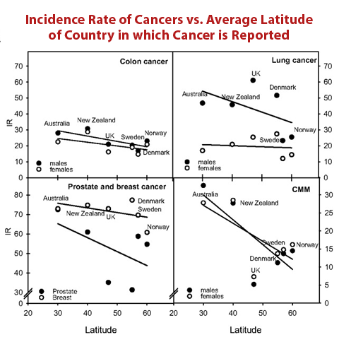
Increased sun exposure does not protect against cancer. In their review, Moan et al showed that proximity to the equator seems to increase the incidence of cancer in at least five of the most common kinds of cancer: colon, lung, prostate, breast, and cutaneous malignant melanoma. When other countries were included in the analysis no effect was found, except in the case of melanoma.

Looking at Karvonen's original study, it is striking to consider that the countries with the world's highest rates of Type I diabetes (in order: Finland, Sweden, Canada, and Norway) also have relatively high rates of vitamin D supplementation. One Finnish study of children born in 1966 found that between 74 and 88% of mothers reported giving their children vitamin D supplements in the form of cod liver oil, typically.3)

Cancer

Main article: Vitamin D and cancer

A number of studies have suggested that sunlight exposure, and the resulting cutaneous synthesis of vitamin D, might have a beneficial influence for certain major cancers, most notably breast, colorectal and prostate cancer. These studies have been based either on using ambient solar UV radiation as a proxy for latitude in those studies looking at the geographical incidence and/or mortality of cancer, or case–control studies using questionnaires in which individuals are asked to recall previous sun exposure. However, this data is not consistent. There are any number of counterexamples in which there is an “inverse latitude gradient.”

Australia and New Zealand vs. Northern Europe

Multiple authors have cited what may be called the “Australia/New Zealand” paradox.4) In his 2006 review,5) Diffey cites “overwhelming evidence that people living in Australia and New Zealand are exposed to considerably more solar UV radiation than those living in northern Europe.” The annual ambient solar UV radiation in southern Australia and New Zealand is about two to three times that in northern Europe (United Kingdom, Ireland, Scandinavia and the Baltic States), rising to more than a fourfold increase in northern Australia. Further, studies of individual exposure to sunlight demonstrate that median doses in Australia are appreciably higher than those measured in a similar cohort of subjects in England.

In the review, Diffey points to epidemiological evidence, which clearly shows that the latitudinal gradient-cancer connection does not hold true at least among a large selection of countries containing large numbers of gentically related people with similar skin types, which is important for the photosynthesis of vitamin D.

Building on Diffey's work, Moan et al, summarize the epidemiological data for the “paradox”:

For some cancers, there may even be a significant inverse latitude gradient. Differences in sun exposure habits and skin types can probably not explain this observation because the inverse relationship remains when we use CMM [cutaneous malignant melanoma] incidence rates as a crude measure for real UV exposures. However, taking more countries into consideration, we see that no reliable north–south gradient can be extracted. There is a large variation of the incidence rates by factors of ≈50 and 5 for prostate cancer and breast cancer, respectively. Even for countries at the same latitude, large differences are found.

Moan et al.6)

United States

In the case of the gradient in different rates of cancer supposedly observed in the United States. Darrell S. Rigel, M.D, president of the American Academy for Dermatology has also argued consistently that no scientific studies exist which prove the statement that low low levels of light exposure lead to increases in cancers and other diseases:

The claim is based on a study that finds that overall cancer rates are higher in the northeast United States, a location with lower sunlight levels than many other places in the country. Those making this claim conclude that since the Northeast has lower UV levels, this is the reason why cancer rates are higher in this region. However, several studies prove this theory is false. These include studies which show that cancer rates are low in the Northern Plains states – areas of the United States that have lower UV levels then the states in the Northeast. Furthermore, several regional studies have shown that the increased levels of cancer observed in the Northeast states are tied to levels of industrial pollutants rather than levels of UV light.

Darrel S. Rigel, MD

Multiple sclerosis

The latitude at which people spend their youth up to the age of about 15 years correlates with later risk of multiple sclerosis, with people who grow up closer to the equator having a lesser risk of disease onset.7) While a number of commentators have blamed vitamin D deficiency, Bains has argued that near-horizon sunshine is at fault.8)

A common precursor for multiple sclerosis is optic neuritis or inflammationThe complex biological response of vascular tissues to harmful stimuli such as pathogens or damaged cells. It is a protective attempt by the organism to remove the injurious stimuli as well as initiate the healing process for the tissue. of the optic nerve. Optic neuritis can be caused by directly looking at the Sun, something that few people willingly do, but inadvertently happens in locations far from the Equator, in which the Sun is often close to the horizon.

In addition to optic neuritis, multiple sclerosis is correlated with several eye symptoms including diplopia (double vision), nystagmus (jerky eye movements), ocular dysmetria (overshooting eye movements), and internuclear opthalmoplegia (lack of coordination between the eyes).

Causation – the elusive grail of epidemiology

Some have argued that the apparent latitude gradient in disease prevalence speaks for itself, but one cannot impute causation simply by looking at a map. In 2003, L.R. Karhausen wrote a paper entitled “Causation: the elusive grail of epidemiology.” In the review, Karhausen rebukes those who would place undue faith in conclusions based on observational epidemiological studies.

Actually, there is no experience of causation: events do not wear their causal credentials on their faces.

L.R. Karhausen 9)

Without a precise understanding of underlying disease mechanisms, researchers could just as well conclude, for example, that sunshine exposure causes teenage pregnancy.

Image from American Human Development Project of the Social Science Research Council. URL: https://measureofamerica.org/maps/

===== Notes and comments =====

  • Legacy content

need a new article? vitamin D production on skin

Trevor talks about this here: https://www.marshallprotocol.com/view_topic.php?id=13471

J Invest Dermatol. 2010 Feb;130(2):546-53. Epub 2009 Oct 8. Vitamin D production after UVB exposure depends on baseline vitamin D and total cholesterol but not on skin pigmentation.

Bogh MK, Schmedes AV, Philipsen PA, Thieden E, Wulf HC. Department of Dermatology, Copenhagen University Hospital, Bispebjerg, Copenhagen NV Vejle, Denmark. bogh@dadlnet.dk Erratum in: J Invest Dermatol. 2010 Jun;130(6):1751. Comment in: J Invest Dermatol. 2010 Feb;130(2):330. J Invest Dermatol. 2010 Aug;130(8):2139-41. J Invest Dermatol. 2010 Feb;130(2):346-8. Abstract UVB radiation increases serum vitamin D level expressed as 25-hydroxyvitamin-DThe vitamin D metabolite widely (and erroneously) considered best indicator of vitamin D "deficiency." Inactivates the Vitamin D Nuclear Receptor. Produced by hydroxylation of vitamin D3 in the liver. (3) (25(OH)D), but the influence of skin pigmentation, baseline 25(OH)D level, and total cholesterol has not been well characterized. To determine the importance of skin pigmentation, baseline 25(OH)D level, and total cholesterol on 25(OH)D production after UVB exposure, 182 persons were screened for 25(OH)D level. A total of 50 participants with a wide range in baseline 25(OH)D levels were selected to define the importance of baseline 25(OH)D level. Of these, 28 non-sun worshippers with limited past sun exposure were used to investigate the influence of skin pigmentation and baseline total cholesterol. The participants had 24% of their skin exposed to UVB (3 standard erythema doses) four times every second or third day. Skin pigmentation and 25(OH)D levels were measured before and after the irradiations. Total cholesterol was measured at baseline. The increase in 25(OH)D level after UVB exposure was negatively correlated with baseline 25(OH)D level (P<0.001) and positively correlated with baseline total cholesterol level (P=0.005), but no significant correlations were found with constitutive or facultative skin pigmentation. In addition, we paired a dark-skinned group with a fair-skinned group according to baseline 25(OH)D levels and found no differences in 25(OH)D increase after identical UVB exposure. PMID: 19812604

J Hum Evol. 2000 Jul;39(1):57-106.

The evolution of human skin coloration.

Jablonski NG, Chaplin G. Department of Anthropology, California Academy of Sciences, Golden Gate Park, San Francisco, CA 94118-4599, USA. njablonski@calacademy.org Abstract Skin color is one of the most conspicuous ways in which humans vary and has been widely used to define human races. Here we present new evidence indicating that variations in skin color are adaptive, and are related to the regulation of ultraviolet (UV) radiation penetration in the integument and its direct and indirect effects on fitness. Using remotely sensed data on UV radiation levels, hypotheses concerning the distribution of the skin colors of indigenous peoples relative to UV levels were tested quantitatively in this study for the first time. The major results of this study are: (1) skin reflectance is strongly correlated with absolute latitude and UV radiation levels. The highest correlation between skin reflectance and UV levels was observed at 545 nm, near the absorption maximum for oxyhemoglobin, suggesting that the main role of melanin pigmentation in humans is regulation of the effects of UV radiation on the contents of cutaneous blood vessels located in the dermis. (2) Predicted skin reflectances deviated little from observed values. (3) In all populations for which skin reflectance data were available for males and females, females were found to be lighter skinned than males. (4) The clinal gradation of skin coloration observed among indigenous peoples is correlated with UV radiation levels and represents a compromise solution to the conflicting physiological requirements of photoprotection and vitamin D synthesis. The earliest members of the hominid lineage probably had a mostly unpigmented or lightly pigmented integument covered with dark black hair, similar to that of the modern chimpanzee. The evolution of a naked, darkly pigmented integument occurred early in the evolution of the genus Homo. A dark epidermis protected sweat glands from UV-induced injury, thus insuring the integrity of somatic thermoregulation. Of greater significance to individual reproductive success was that highly melanized skin protected against UV-induced photolysis of folate (Branda & Eaton, 1978, Science201, 625-626; Jablonski, 1992, Proc. Australas. Soc. Hum. Biol.5, 455-462, 1999, Med. Hypotheses52, 581-582), a metabolite essential for normal development of the embryonic neural tube (Bower & Stanley, 1989, The Medical Journal of Australia150, 613-619; Medical Research Council Vitamin Research Group, 1991, The Lancet338, 31-37) and spermatogenesis (Cosentino et al., 1990, Proc. Natn. Acad. Sci. U.S.A.87, 1431-1435; Mathur et al., 1977, Fertility Sterility28, 1356-1360).As hominids migrated outside of the tropics, varying degrees of depigmentation evolved in order to permit UVB-induced synthesis of previtamin D(3). The lighter color of female skin may be required to permit synthesis of the relatively higher amounts of vitamin D(3)necessary during pregnancy and lactation. Skin coloration in humans is adaptive and labile. Skin pigmentation levels have changed more than once in human evolution. Because of this, skin coloration is of no value in determining phylogenetic relationships among modern human groups. Copyright 2000 Academic Press.

PMID: 10896812

Am J Phys Anthropol. 2009 Aug;139(4):447-50. The evolution of light skin color: role of vitamin D disputed. Robins AH. Department of Medicine, Division of Pharmacology, University of Cape Town Medical School, Observatory, South Africa 7925. ashley.robins@uct.ac.za Comment in: Am J Phys Anthropol. 2009 Aug;139(4):451-61.

PMID: 19425095

  • atherosclerosis in African Americans – Vitamin D is widely used to treat patients with osteoporosis and/or low vitamin D levels based on a medically accepted normal range. This “normal” range is typically applied to all race groups, although it was established predominantly in whites. It is thought that as low vitamin D levels rise to the normal range with supplementation, protection from bone and heart disease (atherosclerosis) may increase, as well. Blacks generally have lower vitamin D levels than whites, partly because their darker skin pigmentation limits the amount of the vitamin produced by sunlight. “Despite” these lower vitamin D levels and dietary calcium ingestion, blacks naturally experience lower rates of osteoporosis and have far less calcium in their arteries. Studies further reveal that black patients with diabetes have half the rate of heart attack as whites, when provided equal access to health care. A 2010 study (explained here) determined the relationship between circulating vitamin D levels and arterial calcium in 340 black men and women with type 2 diabetes.10) The team concluded that higher circulating levels of 25-D in blacks were associated with higher levels of calcified atherosclerotic plaque.

Serum 25(OH)D concentrations in sunny Israel.Saliba W, Rennert HS, Kershenbaum A, Rennert G Osteoporos Int Mar 2011; Full text via publisher | Download citation Affiliation Department of Community Medicine and Epidemiology, Carmel Medical Center, Clalit Health Services, and Bruce Rappaport Faculty of Medicine, Technion-Israel Institute of Technology, 7 Michal St., Haifa, 34362, Israel, saliba_wa@clalit.org.il. Abstract This study assesses vitamin D status in Israel. Serum 25(OH)D levels <25 and <50 nmol/L are common in Israel with noted differences between Arabs and Jews, Arab females were particularly at high risk. These findings may require public health intervention at the population level. INTRODUCTION: Small studies from Israel have suggested a high prevalence of hypovitaminosis D. The objective of this study was to evaluate the extent of hypovitaminosis D among demographic subgroups in Israel. METHODS: The data of this study are from the Clalit Health Services (CHS) which is a non-for-profit health maintenance organization (HMO) covering more than half of the Israeli population. We included all CHS members for whom a 25(OH)D test result in 2009 was available and who were not taking vitamin D supplements in 2008-2009 before that 25(OH)D result. Complete data were available for 198,834 members. RESULTS: The mean level of 25(OH)D was 51.9 ± 24.5 nmol/L and was higher in summer compared to winter (P < 0.0001). Level <25, <37.5, and <50 nmol/L were detected in 14.4%, 30.7%, and 49.9% of tests; 16.4% had levels >75 nmol/L. Females had higher prevalence of 25(OH)D levels < 50 nmol/L which were found in 51.8% of females versus 45.0% in males (P < 0.0001); 76.7% of the Arabs had levels <50 nmol/L versus 46.5% in Jews (P < 0.0001). Arabs females were particularly at high risk for 25(OH)D <50 nmol/L; 84.8% of them had levels <50 nmol/L versus 48.1% of Jewish females (P < 0.0001). The relation of 25(OH)D levels with age had a sinusoidal shape among Jews, a U-shape in Arab females, and inverse linear pattern in Arab males. CONCLUSIONS: 25(OH)D levels <25 and <50 nmol/L are common in Israel. Public health measures are needed for values lesser than about 30 nmol/L and further monitoring of concentrations between about 30 and 50 nmol/L to determine if there are adverse health effects.

===== References =====

1)
Mohr SB, Garland CF, Gorham ED, Garland FC. The association between ultraviolet B irradiance, vitamin D status and incidence rates of type 1 diabetes in 51 regions worldwide. Diabetologia. 2008 Aug;51(8):1391-8. doi: 10.1007/s00125-008-1061-5. Epub 2008 Jun 12.
[PMID: 18548227] [DOI: 10.1007/s00125-008-1061-5]
2)
Karvonen M, Viik-Kajander M, Moltchanova E, Libman I, LaPorte R, Tuomilehto J. Incidence of childhood type 1 diabetes worldwide. Diabetes Mondiale (DiaMond) Project Group. Diabetes Care. 2000 Oct;23(10):1516-26. doi: 10.2337/diacare.23.10.1516.
[PMID: 11023146] [DOI: 10.2337/diacare.23.10.1516]
3)
Hyppönen E, Sovio U, Wjst M, Patel S, Pekkanen J, Hartikainen A, Järvelinb M. Infant vitamin d supplementation and allergic conditions in adulthood: northern Finland birth cohort 1966. Ann N Y Acad Sci. 2004 Dec;1037:84-95. doi: 10.1196/annals.1337.013.
[PMID: 15699498] [DOI: 10.1196/annals.1337.013]
4)
Moan J, Porojnicu AC, Dahlback A, Setlow RB. Addressing the health benefits and risks, involving vitamin D or skin cancer, of increased sun exposure. Proc Natl Acad Sci U S A. 2008 Jan 15;105(2):668-73. doi: 10.1073/pnas.0710615105. Epub 2008 Jan 7.
[PMID: 18180454] [PMCID: 2206594] [DOI: 10.1073/pnas.0710615105]
5)
Diffey B. Do we need a revised public health policy on sun exposure?. Br J Dermatol. 2006 Jun;154(6):1046-51. doi: 10.1111/j.1365-2133.2006.07268.x.
[PMID: 16704633] [DOI: 10.1111/j.1365-2133.2006.07268.x]
6)
Drace JE, Pelc NJ. Measurement of skeletal muscle motion in vivo with phase-contrast MR imaging. J Magn Reson Imaging. 1994 Mar-Apr;4(2):157-63. doi: 10.1002/jmri.1880040211.
[PMID: 8180454] [DOI: 10.1002/jmri.1880040211]
7)
Kurtzke JF. Multiple sclerosis in time and space--geographic clues to cause. J Neurovirol. 2000 May;6 Suppl 2:S134-40.
[PMID: 10871801]
8)
Bains W. Exposure of the eyes to near-horizon sunshine may be a trigger for multiple sclerosis. Med Hypotheses. 2010 Mar;74(3):428-32. doi: 10.1016/j.mehy.2009.09.054. Epub 2009 Nov 10.
[PMID: 19906495] [DOI: 10.1016/j.mehy.2009.09.054]
9)
Karhausen LR. Causation: the elusive grail of epidemiology. Med Health Care Philos. 2000;3(1):59-67. doi: 10.1023/a:1009970730507.
[PMID: 11080970] [DOI: 10.1023/a:1009970730507]
10)
Freedman BI, Wagenknecht LE, Hairston KG, Bowden DW, Carr JJ, Hightower RC, Gordon EJ, Xu J, Langefeld CD, Divers J. Vitamin d, adiposity, and calcified atherosclerotic plaque in african-americans. J Clin Endocrinol Metab. 2010 Mar;95(3):1076-83. doi: 10.1210/jc.2009-1797. Epub 2010 Jan 8.
[PMID: 20061416] [PMCID: 2841532] [DOI: 10.1210/jc.2009-1797]
home/pathogenesis/vitamind/latitude.txt · Last modified: 09.14.2022 by 127.0.0.1
© 2015, Autoimmunity Research Foundation. All Rights Reserved.