Home

Inflammatory bowel disease (Crohn's disease and ulcerative colitis)

Introduction

Mayo Clinic overview

The metagenomic approach allowed us to detect a reduced complexity of the bacterial phylum Firmicutes as a signature of the faecal microbiotaThe bacterial community which causes chronic diseases - one which almost certainly includes multiple species and bacterial forms. in patients with CD. It also indicated the presence of new bacterial species. 1)

Gut microbial induction of host immune maturation exemplifies host-microbe mutualism. We colonized germ-free (GF) mice with mouse microbiota (MMb) or human microbiotaThe bacterial community in the human body. Many species in the microbiota contribute to the development of chronic disease. (HMb) to determine whether small intestinal immune maturation depends on a coevolved host-specific microbiota. Gut bacterial numbers and phylum abundance were similar in MMb and HMb mice, but bacterial species differed, especially the Firmicutes. HMb mouse intestines had low levels of CD4(+) and CD8(+) T cells, few proliferating T cells, few dendritic cells, and low antimicrobial peptide expression-all characteristics of GF mice. Rat microbiota also failed to fully expand intestinal T cell numbers in mice. Colonizing GF or HMb mice with mouse-segmented filamentous bacteria (SFB) partially restored T cell numbers, suggesting that SFB and other MMb organisms are required for full immune maturation in mice. Importantly, MMb conferred better protection against Salmonella infection than HMb. A host-specific microbiota appears to be critical for a healthy immune system. 2)

Evidence of infectious cause

These papers are really helpful.3) 4)

  1. Patient response to immunosuppressive medications – Temporary symptomatic remission after taking immunosuppressive medications implies a decrease in the strength of the bacterial die-off reaction known as immunopathologyA temporary increase in disease symptoms experienced by Marshall Protocol patients that results from the release of cytokines and endotoxins as disease-causing bacteria are killed.. That none of these medications make patients better over the long-term is also telling. Several immunosuppressants are used to treat Crohn's including glucocorticoids such as prednisone and budesonide, aminosalicylates such as sulfasalazine and mesalamines, immunomodulators such as azathioprine and methotrexate and biologics such as remicade/infliximab and adalimumab.
  2. Presence of microbes – Bacteria have long been suspected to be the cause of Crohn's Disease. Detection rates for bacterial DNA from L-formsDifficult-to-culture bacteria that lack a cell wall and are not detectable by traditional culturing processes. Sometimes referred to as cell wall deficient bacteria. (a portion of the metagenomic microbiotaThe community of bacterial pathogens including those in an intracellular and biofilm state which cause chronic disease. that cause disease) in Crohn's tissues is variable due to “the fastidious culture requirements of the cell wall-deficient forms.” 5) The following types and species of bacteria have been found in patients with Crohn's:

De Hertogh et al provide a thorough review of the “unidentified persistent pathogen” theory, stating:

Various environmental factors may play a role in the development of CD [Crohn's Disease], but microbes are most consistently implied. This theory is based on epidemiological, clinicopathological, genetic and experimental evidence. 13)

Other researchers have come to similar conclusions:

M. avium subsp paratuberculosis, adherent-invasive E. coli and Candida are good candidates for an infectious aetiology of Crohn's disease on the basis of genetic susceptibility, which relates to impaired function in the defence against intracellular bacteria.

De Chambrunet et al. 14)

The bacterial community, in whole or in part, resident in the bowel of humans is considered to fuel the chronic immune inflammatory conditions characteristic of Crohn's disease and ulcerative colitis. Chronic or recurrent pouchitis in ulcerative colitis patients is responsive to antibiotic therapy, indicating that bacteria are the etiological agents.

Sokol et al. 15)

Bacterial exotoxins downregulate cathelicidin Family of antimicrobial peptides found primarily in immune cells and transcribed by the Vitamin D Receptor. (hCAP-18/LL-37) and human beta-defensin An antimicrobial peptide found primarily in immune cells and transcribed by the Vitamin D Receptor. 1 (HBD-1) expression in the intestinal epithelial cells. 16)

More evidence

PubMed reviews on the microbiology of inflammatory bowel diseases

58. El Zaatari FA, Osato MS, Graham DY. Etiology of Crohn's disease: the role of Mycobacterium avium paratuberculosis. Trends Mol Med. 2001;7:247-252. 59. Harris JE, Lammerding AM. Crohn's disease and Mycobacterium avium subsp. paratuberculosis: current issues. J Food Prot. 2001;64:2103-2110.

Correlation between the immune responses to collagens type I, III, IV and V and Klebsiella pneumoniae in patients with Crohn's disease and ankylosing spondylitis

Crohn's disease

Parasitization of vitreous leukocytes by mollicute-like organisms. Am J Clin Pathol. 1989 Mar;91(3):259-64. Johnson LA, Wirostko E, Wirostko WJ. Crohn's disease uveitis

Uveitis is a symptom, and has many identified causes and associations at this point. Some cases are widely accepted to be due to bacteria or viruses. Other cases are associated with “autoimmune diseases” like Crohns disease and rheumatic arthritis. I suspect that these other cases are due to CWD bacteria. There are several interesting papers that explore this. The researchers found cell-wall deficient bacteria (sometimes called mollicute-like organisms) in the vitreous fluid of patients with sardoidosis, Crohn’s disease, ulcerative colitis, juvenile rheumatoid arthritis, etc.”

Mycobacterium

Aussie-native Dr. Thomas Borody, who was awarded the Marshall Prize in Australia for innovative scientific research and who lectured 3/06 on Long Island, has proposed that Crohn's disease, is caused by Mycobacterium avium paratuberculosis (or MAP for short). The microbe is a distant relative of the tuberculosis and leprosy bacteria.

MP patient reported

Kenc wrote:

This pathogen was suspected in the early part of the last century soon after Crohn's disease was first identified. However, since researchers could not isolate it in Crohn's disease patients they gave up and turned to the concept of Crohn's disease being an immune disorder and so began the big push for immunosuppresive drugs.

Within the last decade there has been a resurgence of interest in MAP. Most Crohn's patients testing positive for MAP (about 45%) have responded well to long term antibiotic therapy in clinical trials (see research by Ira Shafran and by T. Borody). Their response was better than any immunosuppresive drug therapy I know. However, since not all Crohn's patients tested positive for MAP the medical community has remained skeptical of the claim that Crohn's disease is caused by MAP.

I believe the pathogenesis behind the Marshall ProtocolA curative medical treatment for chronic inflammatory disease. Based on the Marshall Pathogenesis. includes the infection by one or more types of bacteria. That would account for the other 55% of Crohn's patients who didn't test positive for MAP. Furthermore the MP should work better than an antibiotic treatment for MAP alone since it would help the immune system to eradicate the other types of bacterium that could be present as well along with MAP.

I tested positive for two other types of bacteria other than MAP. One type of these bacterium comes from tics. I did not take a test for MAP. So, my case is evidence that a Crohn's patient can have non-MAP infections as well.

In spite of the increasing evidence for a microbial cause of Crohn's disease, most (>95% I believe) of the research money available for Crohn's disease, including money from charitable organizations, appears to be directed towards the development of immunosuppressive drugs. The latest craze seems to be for TNF-alpha blockers like Remicade.

The most amazing research I've seen was for MS patients. The idea was to kill all the white blood cells in an MS patient and then use stem cells taken earlier from the patient to repopulate the white blood cells. Of course about half of the patients died. This is modern medical research at its finest! It worked out OK for those that survived. Looking at this with MP eyes, I can see this as a drastic way of getting rid of CWD bacterium in the white blood cells - kill all the cells.

Antibiotics

Researchers at Cedars-Sinai Medical Center have found that a nonabsorbable antibiotic – one that stays in the gut – may by be an effective long-term treatment for irritable bowel syndrome (IBS), a disease affecting more than an estimated 20 percent of Americans. The findings, which showed that participants benefited from the antibiotic use even after the course of treatment ended, support previously published research identifying small intestine bacterial overgrowth (SIBO) as a possible cause of the disease. (Nov. 8, 2005)

“This study is important as it is the first to show that the use of targeted antibiotics results in a more significant and long-lasting improvement in IBS symptoms,” said Mark Pimentel, M.D., director of the GI Motility Program at Cedars-Sinai. “These results clearly show that antibiotics offer a new treatment approach – and a new hope – for people with IBS.”

The randomized, double blind study involved 87 patients. Those on the rifaximin experienced a 37 percent overall improvement of their IBS symptoms as compared to 23 percent on the placebo. Among study subjects whose primary symptom was diarrhea, those on the antibiotic showed more than twice the improvement of those on the placebo (49 percent vs. 23 percent). Patients received the drug (or placebo) for 10 days and were then followed for a total of 10 weeks. Participants kept a stool diary, took a questionnaire and were given methane breath tests. The positive effects of the drug were shown to continue throughout most of the 10-week study, not just during the actual antibiotic course.

Because the cause of IBS has been elusive, treatments for the disease have historically focused on reducing its symptoms – diarrhea and constipation – by giving medications that either slow or speed up the digestive process. In 2000, Pimentel linked bloating, the most common symptom of IBS, to bacterial fermentation, showing that small intestine bacteria overgrowth (SIBO) may be the causative factor in IBS (The American Journal of Gastroenterology, Dec. 2000).

To show evidence of small intestine bacterial overgrowth, participants in both studies were given a lactulose breath test, which monitors the level of hydrogen and methane (the gases emitted by fermented bacteria) on the breath. In the first study, an abnormal breath methane profile was shown to be 100 percent predictive of constipation-predominant IBS. In the current study, the correlation between the amount of methane and the amount of constipation was confirmed, another key finding.

“We were pleased – but not surprised – with the results of this study,” said Pimentel.

Irritable Bowel Syndrome is an intestinal disorder that causes abdominal pain or discomfort, cramping or bloating and diarrhea and constipation. It is a long-term condition that usually begins in adolescence or in early adult life. Episodes may be mild or severe and may be exacerbated by stress. It is one of the top ten most frequently diagnosed conditions among U.S. physicians and affects women more often than men.

Rifaximin is made by Salix Pharmaceuticals, Inc. Funding for the study was provided by Salix Pharmaceuticals, Inc.

One of only five hospitals in California whose nurses have been honored with the prestigious Magnet designation, Cedars-Sinai Medical Center is one of the largest nonprofit academic medical centers in the Western United States. Additional information is available at https://www.cedars-sinai.edu

Targeted Antibiotics Lead to Long-lasting Improvement in IBS Symptoms

It is apparently very rare for a patient to actually be allergic to OlmesartanMedication taken regularly by patients on the Marshall Protocol for its ability to activate the Vitamin D Receptor. Also known by the trade name Benicar. (see Research below). An alternative approach where there is present a confirmed mycobacterial infection was recorded here MAP Abx .

Antimicrobial peptides

Explaining how the antimicrobial peptidesBody’s naturally produced broad-spectrum antibacterials which target pathogens. (the body's own antibiotics) are employed against the pathogens which cause Crohn's disease.

The VDRThe Vitamin D Receptor. A nuclear receptor located throughout the body that plays a key role in the innate immune response. is responsible for the Cathelicin Anti-Microbial Peptides. It is also responsible for transcribing the genes of the Beta Defensins.

This presentation explains how those, and the other Defensins, are important, and I think that those of you who track the science will find it very interesting indeed.

The presentation runs for 30 minutes. Crohns

Serum levels of 'vitamin' D

BACKGROUND: The active form of vitamin D (1,25(OH)2D3) has been shown to inhibit development of inflammatory bowel disease (IBD) in IL-10 KO mice.

Data point to a critical role for the VDR and 1,25(OH)2D3 in control of innate immunityThe body's first line of defense against intracellular and other pathogens. According to the Marshall Pathogenesis the innate immune system becomes disabled as patients develop chronic disease. and the response of the colon to chemical injury. 17)

Department of Internal Medicine, University of Oulu, Finland. OBJECTIVES. To explore the relationships between vitamin D intake, serum parathyroid hormone (PTH) and 25-hydroxyvitamin D (250HD) concentrations, and bone mineral density (BMD) in inflammatory bowel disease (IBD). SUBJECTS. One hundred and fifty randomly selected patients with IBD from the hospital register and 73 healthy controls.

Patients with IBD have lower serum levels of 250HD than healthy controls, but similar serum PTH concentrations and vitamin D intake. Vitamin D intake, and the serum levels of 250HD and PTH are not associated with BMD, and malabsorption is unlikely to be a major factor in the aetiology of bone loss in unselected IBD patients. 18)

Culture independent analysis of ileal mucosa reveals a selective increase in invasive Escherichia coli of novel phylogeny relative to depletion of Clostridiales in Crohn's disease involving the ileum.

The number of E. coli in situ correlated with the severity of ileal disease (rho 0.621, P<0.001) and invasive E. coli was restricted to inflamed mucosa. E. coli strains isolated from the ileum were predominantly novel in phylogeny, displayed pathogen-like behavior in vitroA technique of performing a given procedure in a controlled environment outside of a living organism - usually a laboratory. and harbored chromosomal and episomal elements similar to those described in extraintestinal pathogenic E. coli and pathogenic Enterobacteriaceae. These data establish that dysbiosis of the ileal mucosa-associated flora correlates with an ileal Crohn's disease (ICD) phenotype, and raise the possibility that a selective increase in a novel group of invasive E. coli is involved in the etiopathogenesis to Crohn's disease involving the ileum. 19)

A study suggests that the bacteria-immune system 'fight' continues after the instigator bacteria have been cleared by the body, according to Andrew Gewirtz. The fight can result in metabolic syndrome, an important factor in obesity, or inflammatory bowel disease (IBD).

“The implication at present is that it is very important to control the early environment,” Professor Gewirtz said. “We need to examine how this can be achieved – perhaps via breastfeeding, a more diverse diet, probiotics are possibilities.”

Metabolic syndrome involves risk factors, including obesity, which can lead to cardiovascular disease, diabetes and stroke. According to the American Heart Association, about 35 percent of adults are affected by this syndrome.

IBD, which includes Crohn's disease and ulcerative colitis, happens when the intestines become inflamed, leading to abdominal cramps and pain, diarrhea, weight loss and bleeding.

“It is increasingly apparent that bacteria are playing a role in healthy development, and need to be properly managed by the mucosal immune system to avoid inflammatory diseases” Gewirtz explained. 20)

Patient interviews

<html> <div class=“patientinterviews”>

<html> <div class=“patientinterviewboxl”> <div class=“patientinterviewimage”></html><html></div> <div class=“patientinterviewtext”> <div class=“patientinterviewname”></html>Melinda Stiles<html></div></html>

Lyme disease, irritable bowel syndrome/ulcerative colitis, radiculitis

Read the interview

<html></div></div>

<br clear=“left” />

</div> </html>

Interviews of patients with other diseases are also available.

Research

Findings suggest that neither olmesartan nor other ARBs were associated with diarrhea among patients undergoing endoscopy. The spruelike enteropathy recently associated with olmesartan is likely a rare adverse effect and milder presentations are unlikely. 21)

In a methotrexate-induced model of intestinal mucositis, olmesartan reduced inflammationThe complex biological response of vascular tissues to harmful stimuli such as pathogens or damaged cells. It is a protective attempt by the organism to remove the injurious stimuli as well as initiate the healing process for the tissue. and induced enteropathy characterized by severe diarrhea, weight loss, and reduced sucrose activity. 22)

Development, validation and implementation of an in vitro model for the study of metabolic and immune function in normal and inflamed human colonic epithelium. 23)

It is now known that epithelial cells have the capacity to secrete and respond to a range of immunological mediators and this suggests that these cells play a prominent role in the pathogenesis of IBD. Current knowledge about the intestinal epithelium has mainly been obtained using models based on animal cells, transformed human intestinal cell lines and isolated cells from resected colonic bowel segments. Species difference, malignant origin and confounders related to surgery, obviously make these cell models however less applicable for patophysiological studies.

Using a functional peptide cleavage assay it was shown that epithelial cells secreted proteolytic active enzymes and that the functional MMP activity was increased in inflamed IBD mucosa.

Our results revealed that colonic epithelial cells express TLR9, a key pattern recognition receptor. Interestingly, the differentiated epithelial cells, which have been exposed to the luminal bacterial flora in vivoA type of scientific study that analyzes an organism in its natural living environment., were unresponsive to TLR9 ligand stimulation, contrasting findings in the epithelial cell line HT-29 that is cultured continuously in bacteria free environment.

'Western'-style diets, high in fat/sugar, low in fibre, decrease beneficial Firmicutes that metabolise dietary plant-derived polysaccharides to SCFAs and increase mucosa-associated Proteobacteria (including enteric pathogens). 24)

Butyric acid-producing anaerobic bacteria as a novel probiotic treatment approach for inflammatory bowel disease. microbiology research

Butyric acid in functional constipation 25)

Butyric acid in irritable bowel syndrome 26)

Roberts doesn’t know how these gut bacteria could have gotten into the brain. They may have crossed from blood vessels, traveling up nerves from the gut, or even come in through the nose

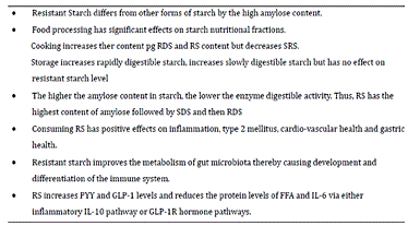
Resistant Starch Regulates Gut Microbiota: Structure, Biochemistry and Cell Signalling

immune mechanisms and interactions between cells of the immune system and tissue environment 27)

Table 1 Yang et al

===== Notes and comments =====

28)

broken link https://mrw.interscience.wiley.com/cochrane/clsysrev/articles/CD007735/frame.html another, maybe thorough review

<DiseaseHierarchy>

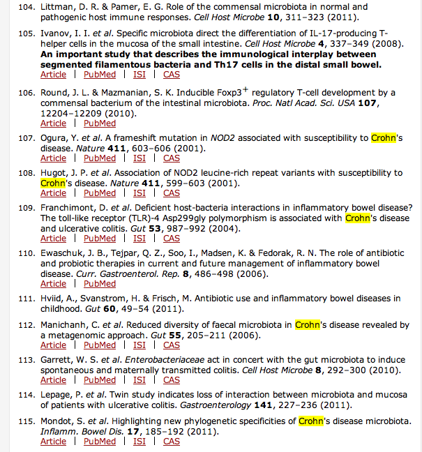
The colon microbiota and inflammatory bowel disease. The microbiome is essential for the activation of host immune responses104. For example, Th17 cell differentiation in the mouse lamina propria requires the presence of segmented filamentous bacteria (SFB)105, and polysaccharide A produced by Bacteroides fragilis mediates the conversion of CD4+ T cells into regulatory T cells106. The inflammatory bowel diseases have long been considered to reflect interactions between microbes and the host. IBD susceptibility is associated with host polymorphisms in bacterial sensor genes such as nucleotide-binding oligomerization domain-containing 2 (NOD2; also known as caspase recruitment domain-containing protein 15 (CARD15))107, 108 and Toll-like receptor 4 (TLR4)109, and symptoms in patients with IBD sometimes improve following antibiotic treatment110. Early childhood exposure to antibiotics has been associated with a significantly increased risk for Crohn's disease111, suggesting that gut microbiome perturbations are important for disease risk. Microbial diversity is significantly diminished in Crohn's disease112, suggesting a decreased gut microbiome resilience that could affect immune interactions. Gut microbiome population structures in patients with ulcerative colitis or Crohn's disease19 depart from normality, but remain clustered by disease within their characteristic deviated patterns. Specific bacteria of the Enterobacteriaceae family may act together with a disordered microbiome to increase the risk of ulcerative colitis113. Between twins that are discordant for ulcerative colitis, those affected had significantly reduced bacterial diversity, but increased proportions of Actinobacteria and Proteobacteria114. Patients with Crohn's disease have over-representation of Enterococcus faecium and of several Proteobacteria compared with controls115. The microbial patterns observed for the conditions described above are preliminary, and their specificity and causal direction have not been established.

Bacteria-immune system 'fight' can lead to chronic diseases, study suggests ATLANTA – Results from a study conducted at Georgia State University suggest that a “fight” between bacteria normally living in the intestines and the immune system, kicked off by another type of bacteria, may be linked to two types of chronic disease.

The study suggests that the “fight” continues after the instigator bacteria have been cleared by the body, according to Andrew Gewirtz, professor of biology at the GSU Center for Inflammation, Immunity and Infection. That fight can result in metabolic syndrome, an important factor in obesity, or inflammatory bowel disease (IBD).

The results were published in the journal Cell Host & Microbe.

“The implication at present is that it is very important to control the early environment,” Gewirtz said. “We need to examine how this can be achieved – perhaps via breastfeeding, a more diverse diet, probiotics are possibilities.”

The study's results are important as instances of chronic diseases like metabolic syndrome and IBD are increasing rapidly among humans, he explained.

Metabolic syndrome involves risk factors, including obesity, which can lead to cardiovascular disease, diabetes and stroke. According to the American Heart Association, about 35 percent of adults are affected by this syndrome.

IBD, which includes Crohn's disease and ulcerative colitis, happens when the intestines become inflamed, leading to abdominal cramps and pain, diarrhea, weight loss and bleeding.

More than 600,000 Americans annually have some kind of inflammatory bowel disease, according to the American Academy of Family Physicians.

Bacteria normally live in the gut of humans, with the average human having about 4 pounds of bacteria living there.

“It is increasingly apparent that bacteria are playing a role in healthy development, and need to be properly managed by the mucosal immune system to avoid inflammatory diseases” Gewirtz explained.

Cell Host Microbe. 2012 Aug 16;12(2):139-52. Epub 2012 Aug 2. Transient Inability to Manage Proteobacteria Promotes Chronic Gut Inflammation in TLR5-Deficient Mice. Carvalho FA, Koren O, Goodrich JK, Johansson ME, Nalbantoglu I, Aitken JD, Su Y, Chassaing B, Walters WA, González A, Clemente JC, Cullender TC, Barnich N, Darfeuille-Michaud A, Vijay-Kumar M, Knight R, Ley RE, Gewirtz AT. Source Center for Inflammation, Immunity, and Infection and Department of Biology, Georgia State University, Atlanta, GA 30303, USA; Clermont Université, Université d'Auvergne, NEURO-DOL, BP 10448, F-63000, Clermont-Ferrand, France; Inserm, U1107, F-63001 Clermont-Ferrand, France. Abstract Colitis results from breakdown of homeostasis between intestinal microbiota and the mucosal immune system, with both environmental and genetic influencing factors. Flagellin receptor TLR5-deficient mice (T5KO) display elevated intestinal proinflammatory gene expression and colitis with incomplete penetrance, providing a genetically sensitized system to study the contribution of microbiota to driving colitis. Both colitic and noncolitic T5KO exhibited transiently unstable microbiotas, with lasting differences in colitic T5KO, while their noncolitic siblings stabilized their microbiotas to resemble wild-type mice. Transient high levels of proteobacteria, especially enterobacteria species including E. coli, observed in close proximity to the gut epithelium were a striking feature of colitic microbiota. A Crohn's disease-associated E. coli strain induced chronic colitis in T5KO, which persisted well after the exogenously introduced bacterial species had been eliminated. Thus, an innate immune deficiency can result in unstable gut microbiota associated with low-grade inflammation, and harboring proteobacteria can drive and/or instigate chronic colitis. Copyright © 2012 Elsevier Inc. All rights reserved. PMID: 22863420

Systemic Toll-Like Receptor Ligands Modify B-cell Responses in Human Inflammatory Bowel DiseaseMarie McDonnell, MD,* YanMei Liang, RN,† Ansu Noronha, MD,‡ Jennifer Coukos, BS,‡ Dennis L. Kasper, MD,§ Francis A. Farraye, MD,‡ and Lisa M. Ganley-Leal, PhD† Background: Bacteria have a central, although poorly under- stood, role in inflammatory bowel disease (IBD). Host–bacteria interactions primarily take place in the gastrointestinal tract, but cells may also encounter translocated bacteria in the bloodstream. IBD is associated with activated, circulating Toll-like receptor (TLR)2 and TLR4-expressing B cells suggesting that blood-borne microbial TLR ligands modulate B cell responses. Methods: Serum levels of lipopolysaccharide (LPS)/endotoxin and high mobility group box 1 (HMGB1), an endogenous TLR ligand, were quantified in Crohn’s disease (CD) and ulcerative co- litis (UC). Responses of purified B cells to LPS and HMGB1 were correlated with levels of systemic TLR ligands and clinical parameters of disease. Results: While IBD patients have increased levels of blood LPS, the net effect of endotoxemia has unexpected characteristics illus- trating that LPS has both pro- and antiinflammatory roles through TLR4þ B cells. Experimental treatment of B cells demonstrates that the antiinflammatory effect of LPS is due to its hypo-acyla- tion of lipid A suggesting an increased prevalence of systemic, hypo-acylated LPS in CD. In contrast, high levels of LPS are associated with disease activity in UC. HMGB1 activates B cells through TLR2A receptor which is expressed on the surface of certain cells and recognizes native or foreign substances and passes on appropriate signals to the cell and/or the nervous system. and CD36. Serum levels of HMGB1 correlate with spontaneous IL-8 production by B cells suggesting that blood- borne TLR2 ligands increase B-cell activation in vivo. Conclusions: Systemic TLR ligands modulate B cells towards either proinflammatory or antiinflammatory activity depending on the predominant ligand(s). Further, the circulating B cell may rep- Received for publication June 3, 2010; Accepted June 14, 2010. From the *Section of Endocrinology, Evans Biomedical Research Center, Boston Medical Center, Boston, Massachusetts, †Section of Infectious Disease, Department of Medicine, Boston University School of Medicine, Boston Medical Center, Boston, Massachusetts, ‡Section of Gastroenterology, Boston Medical Center, Boston, Massachusetts, §Channing Laboratory, Department of Medicine, Brigham and Women’s Hospital, Harvard Medical School, Boston, Massachusetts. Supported by Broad Medical Research Program of the Broad Foundation (to L.G.-L.), BD Grant Award 2007 (to L.G.-L.), and a Department of Medicine Pilot Award (to M.M.). Reprints: Lisa M. Ganley-Leal, PhD, Section of Infectious Diseases, Boston University School of Medicine, 650 Albany St., Room 630, Boston, MA 02118 (e-mail: lisa.ganleyleal@bmc.org) Copyright VC 2010 Crohn’s & Colitis Foundation of America, Inc.

DOI 10.1002/ibd.21424

Published online 30 August 2010 in Wiley Online Library

(wileyonlinelibrary.com). 298

resent an important proxy for quantifying the LPS lipid A acyla- tion burden in patients with IBD.

From the above paper: Most expertise in the area of ‘‘Human MicrobiomeThe bacterial community in the human body. Many species in the microbiota contribute to the development of chronic disease.’’ is focused on the flora of the gastrointestinal tract, the largest, most complex microbiome. Our results suggests that it is highly probable that a blood microbiome exists and, in parallel with what is known of the microbiome of other anatomical sites, may be altered

in disease states with important immunologic consequences for health and homeostasis.50

Gastroenterology. 2011 Jul;141(1):227-36. Epub 2011 Apr 16.

Twin study indicates loss of interaction between microbiota and mucosa of patients with ulcerative colitis. Lepage P, Häsler R, Spehlmann ME, Rehman A, Zvirbliene A, Begun A, Ott S, Kupcinskas L, Doré J, Raedler A,Schreiber S.

Source Institute of Clinical Molecular Biology, Christian-Albrechts University-Kiel, Kiel, Germany.

Abstract BACKGROUND &#38; AIMS: Interactions between genetic and environmental factors are believed to be involved in onset and initiation of inflammatory bowel disease. We analyzed the interaction between gastrointestinal mucosal microbiota and host genes in twin pairs discordant for ulcerative colitis (UC) to study the functional interaction between microbiota and mucosal epithelium.

METHODS: Biopsy were collected from sigmoid colon of UC patients and their healthy twins (discordant twin pairs) and from twins without UC. Microbiota profiles were determined from analysis of 16S ribosomal DNA libraries; messenger RNA profiles were determined by microarray analysis.

RESULTS: Patients with UC had dysbiotic microbiota, characterized by less bacterial diversity and more Actinobacteria and Proteobacteria than that of their healthy siblings; healthy siblings from discordant twins had more bacteria from the Lachnospiraceae and Ruminococcaceae families than twins who were both healthy. In twins who were both healthy, 34 mucosal transcripts correlated with bacterial genera, whereas only 25 and 11 correlated with bacteria genera in healthy individuals and their twins with UC, respectively. Transcripts related to oxidative and immune responses were differentially expressed between patients with UC and their healthy twins.

CONCLUSIONS: The transcriptional profile of the mucosa appears to interact with the colonic microbiota; this interaction appears to be lost in colon of patients with UC. Bacterial functions, such as butyrate production, might affect mucosal gene expression. Patients with UC had different gene expression profiles and lower levels of biodiversity than their healthy twins, as well as unusual aerobic bacteria. Patients with UC had lower percentages of potentially protective bacterial species than their healthy twins.

Copyright © 2011 AGA Institute. Published by Elsevier Inc. All rights reserved.

PMID:

21621540

Inflamm Bowel Dis. 2011 Jan;17(1):185-92. doi: 10.1002/ibd.21436. Epub 2010 Aug 18.

Highlighting new phylogenetic specificities of Crohn's disease microbiota. Mondot S, Kang S, Furet JP, Aguirre de Carcer D, McSweeney C, Morrison M, Marteau P, Doré J, Leclerc M.

Source UMR1319 MICALIS, INRA, Domaine de Vilvert, 78350 Jouy en Josas, France. marion.leclerc@jouy.inra.fr

Abstract BACKGROUND: Recent studies suggest that gastrointestinal (GI) microbes play a part in the pathogenesis of Crohn's disease (CD).

METHODS: Fecal samples were collected from 16 healthy individuals and 16 CD patients (age- and sex-matched). The DNA extracted from these samples were subjected to two different methods of microbiome analysis. Specific bacterial groups were quantified by real-time polymerase chain reaction (PCR) methods using primers designed using a high-throughput in-house bioinformatics pipeline. The same DNA extracts were also used to produce fluorescently labeled cRNA amplicons to interrogate a custom-designed phylogenetic microarray for intestinal bacteria.

RESULTS: Even though the intersubject variability was high, differences in the fecal microbiomes of healthy and CD patients were detected. Faecalibacterium prausnitzii and Escherichia coli were more represented in healthy and ileal CD patients, respectively. Additionally, probes specific for Ruminococcus bromii, Oscillibacter valericigenes, Bifidobacterium bifidum, and Eubacterium rectale produced stronger hybridization signals with the DNA samples from healthy subjects. Conversely, species overrepresented in CD patients were E. coli, Enterococcus faecium, and species from the Proteobacteria not normally found in the healthy human GI tract. Furthermore, we detected “healthy specific” molecular species or operational taxonomic units (OTUs) that are not closely related to any known species (Faecalibacterium, Subdoligranulum, and Oscillospora species), indicating that the phylogenetic dysbiosis is broader than at strain or species level.

CONCLUSIONS: These two techniques of microbiome analysis provided a statistically robust new picture of the dysbiosis in fecal microbiota from ileal CD patients. Specifically, we identified a set of six species discriminant for CD, which provides a preliminary diagnostic tool.

Copyright © 2010 Crohn's & Crohn's & Colitis Foundation of America, Inc.

PMID:

20722058

Wang, T.-T., B. Dabbas, et al. (2010). “Direct and Indirect Induction by 1,25-Dihydroxyvitamin D3 of the NOD2/CARD15-Defensin Œ≤2 Innate Immune Pathway Defective in Crohn Disease.” Journal of Biological Chemistry 285(4): 2227-2231.

Wang et al have done it again (attached).

This time figuring out the link between VDR and expression of Beta-defensin-2. In the process figuring out why the NOD gene is so important to Crohn's …

Note their emerging recognition of the importance of the VDR in the innate immune system… All they need to recognize now is that the VDR is knocked out by persistent pathogens…

..Trevor..

Vitamin D signaling through its nuclear vitamin D receptorA nuclear receptor located throughout the body that plays a key role in the innate immune response. has emerged as a key regulator of innate immunity in humans. Here we show that hormonal vitamin D, 1,25-dihydroxyvitamin DPrimary biologically active vitamin D hormone. Activates the vitamin D nuclear receptor. Produced by hydroxylation of 25-D. Also known as 1,25-dihydroxycholecalciferol, 1,25-hydroxyvitamin D and calcitirol., robustly stimulates expression of pattern recognition receptor NOD2/CARD15/IBD1 gene and protein in primary human monocytic and epithelial cells. The vitamin D receptor signals through distal enhancers in the gene, whose function was validated by chromatin immunoprecipitation and chromatin conformation capture assays. A key downstream signaling consequence of NOD2 activation by agonist muramyl dipeptide is stimulation of NF-κB transcription factor function, which induces expression of the gene encoding antimicrobial peptide defensin β2 (DEFB2/HBD2). Pretreatment with 1,25-dihydroxyvitamin D synergistically induced NF-κB function and expression of genes encoding DEFB2/HBD2 and antimicrobial peptide cathelicidin in the presence of muramyl dipeptide. Importantly, this synergistic response was also seen in macrophages from a donor wild type for NOD2 but was absent in macrophages from patients with Crohn disease homozygous for non-functional NOD2 variants. These studies provide strong molecular links between vitamin D deficiency and the genetics of Crohn disease, a chronic incurable inflammatory bowel condition, as Crohn's pathogenesis is associated with attenuated NOD2 or DEFB2/HBD2 function.

https://www.nejm.org/doi/full/10.1056/NEJMra1102942

Ulcerative Colitis Silvio Danese, M.D., and Claudio Fiocchi, M.D. N Engl J Med 2011; 365:1713-1725November 3, 2011 ArticleReferences Ulcerative colitis was first described in the mid-1800s,1 whereas Crohn's disease was first reported later, in 1932, as “regional ileitis.”2 Because Crohn's disease can involve the colon and shares clinical manifestations with ulcerative colitis, these entities have often been conflated and diagnosed as inflammatory bowel disease, although they are clearly distinct pathophysiological entities. Ulcerative colitis is the most common form of inflammatory bowel disease worldwide. In contrast to Crohn's disease, ulcerative colitis is a disease of the mucosa that is less prone to complications and can be cured by means of colectomy, and in many patients, its course is mild.3 The literature on the pathogenesis and treatment of so-called inflammatory bowel disease has tended to focus on Crohn's disease,4-7 and few articles expressly discuss ulcerative colitis.8,9 Here we review our current understanding of the pathophysiology, diagnosis, and treatment of ulcerative colitis to date; we also compare ulcerative colitis with Crohn's disease and refer to both as inflammatory bowel disease according to the context. PATHOGENESIS Epidemiologic Features Ulcerative colitis and Crohn's disease are disorders of modern society, and their frequency in developed countries has been increasing since the mid-20th century. When inflammatory bowel disease is identified in a new population, ulcerative colitis invariably precedes Crohn's disease and has a higher incidence. The incidence of ulcerative colitis is 1.2 to 20.3 cases per 100,000 persons per year, and its prevalence is 7.6 to 246.0 cases per 100,000 per year, as compared with an incidence of 0.03 to 15.6 cases and a prevalence of 3.6 to 214.0 cases per 100,000 per year for Crohn's disease.10 Among children, however, ulcerative colitis is less prevalent than Crohn's disease.11 The highest incidence and prevalence of inflammatory bowel disease are seen in the populations of Northern Europe and North America and the lowest in continental Asia, where ulcerative colitis is by far the most common form of inflammatory bowel disease.12 A westernized environment and lifestyle is linked to the appearance of inflammatory bowel disease, which is associated with smoking, diets high in fat and sugar, medication use, stress, and high socioeconomic status.13 Inflammatory bowel disease has also been associated with appendectomy.13 Of these factors, only cigarette smoking and appendectomy are reproducibly linked to ulcerative colitis. Smoking is associated with milder disease, fewer hospitalizations, and a reduced need for medications.14 Removal of an inflamed appendix in early life is associated with a decreased incidence of ulcerative colitis,15 whereas the opposite is true for Crohn's disease. Genetic Features The discovery that NOD2 variants are associated with susceptibility to Crohn's disease opened a new era in the study of the genetic basis of inflammatory bowel disease.16,17 In studies of twins, there is stronger concordance with Crohn's disease than with ulcerative colitis, and the identification of a large number of susceptibility loci for Crohn's disease in early genomewide association studies suggested that genetic influences play a greater role in Crohn's disease than in ulcerative colitis.18 A meta-analysis of six such studies recently confirmed the presence of 47 loci associated with ulcerative colitis, of which 19 are specific for ulcerative colitis and 28 are shared with Crohn's disease.19 Several pathways potentially associated with ulcerative colitis were identified in the meta-analysis and in individual studies based on validated loci or chromosomal regions.20 Risk loci for ECM1, HNF4A, CDH1, and LAMB1 implicate dysfunction of the epithelial barrier; an association with DAP suggests a link to apoptosis and autophagy; and associations with PRDM1, IRF5, and NKX2-3 suggest defects in transcriptional regulation. In addition, multiple genes in the interleukin-23 signaling pathway overlap in ulcerative colitis and Crohn's disease (e.g., IL23R, JAK2, STAT3, IL12B, and PTPN2). Several risk loci linked to other immune system–mediated diseases are associated with ulcerative colitis, particularly HLA-DR and genes involved in helper T-cell types 1 and 17 (Th1 and Th17) differentiation, such as IL10, IL7R, IL23R, and IFN-γ. Altogether, genetic studies indicate that both specific and nonspecific gene variants are associated with ulcerative colitis, and the two forms of inflammatory bowel disease share disease pathways. Ulcerative colitis appears to be as genetically heterogeneous as Crohn's disease, but given the large number of implicated genes and the small additive effect of each, genetic screening is not currently indicated to assess the risk of ulcerative colitis. Microbiologic Features Health depends on a beneficial host–microbe interaction. This is certainly true for intestinal health, particularly in the colon, which harbors a greater and more diverse number of microorganisms than any other organ.21 The gut immune system is generally tolerant of this microbial load, and a breakdown in tolerance is postulated to be central to the pathogenesis of inflammatory bowel disease.22 Although loss of tolerance to the gut microbiota is demonstrable in animal models of inflammatory bowel disease, there is only limited evidence for this finding in patients with Crohn's disease and none in those with ulcerative colitis. It has also been postulated that alterations in the composition of the gut microbiota, defects in mucosal immunity, or the two factors combined could lead to ulcerative colitis; however, supportive evidence is sparse. A key issue is the characterization of the gut microbiota in the normal intestine and in the intestine in patients with inflammatory bowel disease. This issue awaits answers from the Human Microbiome Project, which aims to define the composition of the intestinal microbiota in conditions of health and disease.23 There is a consensus that the density of microbiota is greater in patients with ulcerative colitis or Crohn's disease than in healthy control subjects, but whether there are reproducible, disease-specific alterations is unclear.24 The fact that antibiotic therapy has no clinical effect on ulcerative colitis argues against an important role of bacteria in this disease, whereas antibiotics do provide some benefit in luminal Crohn's disease.25 Although serum antibacterial antibodies are present in patients with ulcerative colitis, they are much more common and are found in higher titers in patients with Crohn's disease. Furthermore, the range of antibodies against bacterial antigens (anti-I2, anti-OmpC, and anti-CBir1 antibodies) and fungal antigens (anti–Saccharomyces cerevisiae antibodies [ASCA]) is broader in Crohn's disease, whereas the only ulcerative colitis–associated antibody is perinuclear antineutrophil cytoplasmic antibody (pANCA), which recognizes nuclear antigens that may cross-react with bacterial antigens.26 Mucosal Immune Response Intestinal homeostasis requires a controlled innate immune responseThe body's first line of defense against intracellular and other pathogens. According to the Marshall Pathogenesis the innate immune system becomes disabled as patients develop chronic disease. to the microbiota, which is recognized by toll-like receptors (TLRs) and nucleotide-binding oligomerization domain (NOD)–like receptors on epithelial and immune cells.27 This recognition process contributes to tolerance, but when the process is dysregulated, inflammation ensues. At present, there is no clear evidence of specific, innate immune defects in ulcerative colitis; an increased expression of TLR2 and TLR4 by colonocytes28 is probably secondary to inflammation. In contrast, in Crohn's disease, abnormalities of innate immunity are linked to variants of the NOD2, ATG16L1, and IRGM genes, the products of which normally mediate microbial recognition.16,17,29,30 The production of proinflammatory cytokinesAny of various protein molecules secreted by cells of the immune system that serve to regulate the immune system., such as interleukin-1β, interleukin-6, tumor necrosis factor α (TNF-α), and tumor necrosis factor–like ligand 1 (TL1A), is universally increased in patients with inflammatory bowel disease but does not allow one to discriminate between ulcerative colitis and Crohn's disease. Abnormalities in humoral and cellular adaptive immunity occur in ulcerative colitis. Elevated IgM, IgA, and IgG levels are common in inflammatory bowel disease, but there is a disproportionate increase in IgG1 antibodies in ulcerative colitis.31 Abnormalities of adaptive immunity that differentiate ulcerative colitis from Crohn's disease are defined by mucosal CD4+ T cells, which were initially divided into two lineages: Th1 and type 2 helper T cells (Th2). Crohn's disease is a Th1-like condition, on the basis of evidence of increased production of interferon-γ.32 In contrast, ulcerative colitis represents an atypical Th2 response, as indicated by the presence of nonclassical natural killer T cells in the colon that secrete abundant interleukin-13, which mediates epithelial-cell cytotoxicity, apoptosis, and epithelial-barrier dysfunction.33,34 Interleukin-5–producing Th2-polarized T cells are also present in ulcerative colitis. The balance between Th1 and Th2 has been used to differentiate between ulcerative colitis and Crohn's disease. However, additional helper-cell lineages have recently been delineated, including Th17 cells that produce the proinflammatory cytokineAny of various protein molecules secreted by cells of the immune system that serve to regulate the immune system. interleukin-17, the levels of which are increased in the mucosa of patients with inflammatory bowel disease.35 However, defects in T-cell regulatory function have not been reported in ulcerative colitis.36 Epithelial Cells and Autoimmunity Because inflammation in ulcerative colitis typically does not extend into the small intestine and occurs in proximity to the epithelium, colonocytes are implicated in the pathogenesis of this disease. It has been proposed that the epithelium is diffusely abnormal, irrespective of inflammation.37 Other reported abnormalities in ulcerative colitis include an epithelial-barrier defect and impaired expression of peroxisome proliferator–activated receptor γ (PPAR-γ), a nuclear receptorIntracellular receptor proteins that bind to hydrophobic signal molecules (such as steroid and thyroid hormones) or intracellular metabolites and are thus activated to bind to specific DNA sequences which affects transcription. that regulates inflammatory genes.38 In both ulcerative colitis and Crohn's disease, epithelial cells have a decreased ability to activate suppressor CD8+ T cells, but this abnormality is probably secondary to other immune events.39 Variants of the XPB1 gene, the product of which is a component of the stress response of the endoplasmic reticulum in epithelial cells, have been linked to inflammatory bowel disease, reinforcing the concept that colonocytes are involved in its pathogenesis.40 Autoimmunity may play a role in ulcerative colitis. In addition to pANCA, this disease is characterized by circulating IgG1 antibodies against a colonic epithelial antigen that is shared with the skin, eye, joints, and biliary epithelium41; since these are the sites of extraintestinal manifestations in ulcerative colitis, it is possible that cross-reacting antibodies against the colon cause organ-specific damage. Tropomyosin 5, a structural protein, is the putative target autoantigen of the IgG1 antibodies,42 but evidence of classical antibody-mediated autoimmunity in ulcerative colitis is still lacking. Figure 1FIGURE 1 Current Concepts Concerning the Pathogenesis of Ulcerative Colitis. summarizes our current understanding of the pathogenesis of ulcerative colitis. CLINICAL MANIFESTATIONS Symptoms, Clinical Course, and Assessment of Disease Activity Bloody diarrhea with or without mucus is the hallmark of ulcerative colitis. The onset is typically gradual, often followed by periods of spontaneous remissionTheory that diseases go away of their own accord. and subsequent relapses. Active disease is manifested as mucosal inflammation commencing in the rectum (proctitis) and in some cases spreading to the rest of the colon (Fig. 1A in the Supplementary Appendix, available with the full text of this article at NEJM.org). Although proctitis is frequently associated with fecal urgency and the passage of fresh blood, constipation may paradoxically occur. Proctosigmoiditis, left-sided colitis, extensive colitis, or pancolitis (Fig. 1B in the Supplementary Appendix) may lead to diarrhea, frequent evacuations of blood and mucus, urgency or tenesmus, abdominal pain, fever, malaise, and weight loss, depending on the extent and severity of the disease.43 A small area of inflammation surrounding the appendiceal orifice (cecal patch) can be identified in patients with left-sided ulcerative colitis and in those with proctitis or proctosigmoiditis44 (Fig. 1C in the Supplementary Appendix), but this finding is not specific. The prognosis for patients with ulcerative colitis is generally good during the first decade after diagnosis, with a low rate of colectomy; over time, remission occurs in most patients.3 Assessment of the clinical activity of ulcerative colitis helps the clinician choose diagnostic tests and make therapeutic decisions. Various indexes of disease activity have been developed on the basis of clinical, laboratory, and endoscopic findings, but they are used primarily in clinical trials.45 Complications Acute complications, such as severe bleeding and toxic megacolon (Fig. 1D in the Supplementary Appendix), may occur in patients with extensive or severe inflammation; other problems, such as epithelial dysplasia or cancer, may emerge during the chronic phase (Fig. 1E in the Supplementary Appendix). On the basis of data from referral centers, the cumulative risk of colorectal cancer among patients with chronic ulcerative colitis may reach 20 to 30% at 30 years,46 but the incidence rate is much lower in population-based series (approximately 2%).47 Risk factors for cancer include a long duration of disease, regardless of clinical activity; extensive involvement; a young age at onset; severe inflammation; the presence of primary sclerosing cholangitis; and a family history of colorectal cancer. Although surveillance colonoscopy is recommended for patients at risk, there is no clear evidence that such surveillance increases survival.48 Extraintestinal manifestations involving various organs and systems (e.g., joints, skin, liver, eye, mouth, and coagulation) that either precede the onset of symptoms or appear and evolve in parallel with intestinal manifestations occur in 10 to 30% of patients with ulcerative colitis (Table 1TABLE 1 Complications in Ulcerative Colitis. ). DIAGNOSIS An accurate diagnosis of ulcerative colitis involves defining the extent and severity of inflammation, and this information provides the basis for selecting the most appropriate treatment and for predicting the patient's prognosis. Both endoscopy and biopsy are required to determine specific histologic characteristics; radiologic and ultrasonographic examinations are not critical but may be useful.43 All these investigations aid in differentiating ulcerative colitis from other conditions that have similar presentations (Table 2TABLE 2 Most Common Differential Diagnosis in Ulcerative Colitis. ). Endoscopic Studies Colonoscopy shows a uniformly inflamed mucosa that starts at the anorectal verge and extends proximally, with an abrupt or a gradual transition from affected to normal mucosa. In mild ulcerative colitis, the mucosa has a granular, erythematous appearance, with friability and loss of the vascular pattern. In moderate disease, erosions or microulcerations are evident, whereas in severe ulcerative colitis, shallow ulcerations with spontaneous bleeding are generally seen (Figure 2A, 2B, and 2C in the Supplementary Appendix). In pancolitis, inflammation stops at the ileocecal valve, with occasional limited involvement of the distal ileum, a condition known as backwash ileitis.49 Colonoscopy helps to differentiate ulcerative colitis from Crohn's disease, which is typically characterized by rectal sparing, aphthous ulcers, skip lesions (areas of inflammation alternating with normal mucosa), a cobblestone pattern, and longitudinal, irregular ulcers. In patients with cycles of inflammation and healing and in those with chronic, unremitting inflammation, colonoscopy may reveal pseudopolyps or mucosal bridging (Fig. 2D and 2E in the Supplementary Appendix). If a stricture is detected, multiple biopsies are mandatory to rule out malignant disease; biopsies are also required for surveillance of dysplasia in patients who have the disease for longer than 8 years. Although there is no clear evidence that surveillance prolongs survival,50 biopsy specimens should be taken from all colonic segments, regardless of whether they are inflamed, with a particular focus on irregular mucosa, polypoid lesions, and any raised, dysplasia-associated lesion or mass51 (Fig. 2F in the Supplementary Appendix). Newer endoscopic techniques that are gaining acceptance, such as chromoendoscopy, narrow-band imaging, and autofluorescence imaging, may better delineate suspicious mucosal patterns and improve the detection of dysplasia (Fig. 2G in the Supplementary Appendix).45,52,53 Histologic Evaluation In ulcerative colitis, inflammation is characteristically restricted to the mucosal layer, with infiltrates varying in density and composition during active disease or stages of remission (Fig. 3A and 3B in the Supplementary Appendix). Infiltrates consist primarily of lymphocytes, plasma cells, and granulocytes; the last are being particularly prominent during acute flare-ups and accumulate in crypt abscesses54 (Fig. 3C in the Supplementary Appendix). Other typical features include goblet-cell depletion, distorted crypt architecture, diminished crypt density, and ulcerations. However, epithelioid granulomas, which are typical of Crohn's disease, are not present. Looking for epithelial dysplasia is critical, given the risk of cancer in patients with long-standing ulcerative colitis; however, dysplasia can occur at any stage without indicating malignant transformation (Fig. 3D in the Supplementary Appendix). There are no exact criteria for the diagnosis of ulcerative colitis, but in most cases, the presence of two or three of the aforementioned histologic features will suffice.55 The severity of inflammation on histologic examination and the severity of disease on endoscopic examination may not coincide; for instance, histologic findings may indicate severe disease even in a patient with endoscopically quiescent disease. Laboratory Tests Although not diagnostic, laboratory measurements are helpful in assessing and monitoring disease activity and in differentiating ulcerative colitis from other forms of colitis. Blood counts and measurements of the erythrocyte sedimentation rate and the level of fecal lactoferrin or calprotectin help determine the severity of the inflammation. Stool cultures for Clostridium difficile, campylobacter species, and Escherichia coli 0157:H7 are recommended to rule out an infectious cause or complication. Patients with severe, refractory disease should be assessed for cytomegalovirus infection by means of histologic, immunochemical, serologic, culture, or DNA testing.56 A positive test for ASCA or pANCA is not diagnostic, given the limited sensitivity and specificity of the tests; when they are performed in combination, however, the results may help differentiate among ulcerative colitis, Crohn's disease, and indeterminate colitis.57 This last condition affects approximately 10% of patients, who have colitis with features of both Crohn's disease and ulcerative colitis. THERAPY Medical Therapy According to current consensus-based guidelines, the choice of treatment for patients with ulcerative colitis should take into consideration the level of clinical activity (mild, moderate, or severe) combined with the extent of disease (proctitis, left-sided disease, extensive disease, or pancolitis), the course of the disease during follow-up, and patients' preferences.43,58,59 Induction of Remission Sulfasalazine and 5-aminosalicylates (mesalamine, olsalazine, and balsalazide), given orally, rectally (by means of suppository or enema), or both, represent first-line treatment for ulcerative colitis, with an expected remission rate of about 50%. 60 Mild-to-moderate proctitis can be treated with mesalamine suppositories (1 g per day) or enemas (2 to 4 g per day); clinical remission occurs in most patients within 2 weeks, with repeated treatments as needed. If this fails, 5-aminosalicylate enemas (2 to 4 g per day) or glucocorticoid enemas (hydrocortisone at a dose of 100 mg per day, or new preparations such as budesonide or beclomethasone) are a next step.61-63 Patients who do not have a response to rectally administered agents may be given oral glucocorticoids (up to 40 mg of prednisone or its equivalent). Mild-to-moderate left-sided colitis to extensive ulcerative colitis is initially best treated with a combination of rectal and oral 5-aminosalicylate (up to 4.8 g per day).64 Escalating doses of oral 5-aminosalicylate generally produce the best clinical response,65 and a once-daily dose of 5-aminosalicylate (2 g per day) or newer, controlled-release formulations, such as multimatrix 5-aminosalicylate (2.4 g per day) are reported to be as effective and to promote adherence to treatment.66,67 Patients with mild-to-moderate ulcerative colitis that is refractory to rectal therapies and to oral 5-aminosalicylate are candidates for oral glucocorticoids or immunosuppressive agents (azathioprine or 6-mercaptopurine); those who do not have a response to maximal doses of 5-aminosalicylate or oral glucocorticoids should be given intravenous glucocorticoids.59 For patients who continue to require glucocorticoid therapy and for those who do not have a response to it, a good therapeutic option appears to be infliximab, a monoclonal antibody against TNF-α, administered at a dose of 5 mg per kilogram of body weight at 0, 2, and 6 weeks.68 Infliximab in combination with azathioprine (2.5 mg per kilogram) was reported to be superior to infliximab or azathioprine monotherapy for inducing glucocorticoid-free remission in patients with moderate-to-severe ulcerative colitis.69 Many specialists suggest that patients with extensive, severe disease receive a 5-day to 7-day course of intravenous glucocorticoids59; if the disease is unresponsive, then intravenous cyclosporine (2 mg per kilogram) or infliximab is usually the next step. Although cyclosporine can be effective, it generally delays rather than prevents subsequent colectomy70; furthermore, infliximab is increasingly used as an alternative treatment for patients with refractory disease, given its effectiveness and better short-term safety profile as compared with other therapies.71,72 Several schema have been proposed for determining whether colectomy should be performed in patients with unresponsive disease; these schema incorporate stool frequency; levels of C-reactive protein, albumin, and fecal calprotectin; and radiologic and endoscopic findings.73 Composite indexes appear to be the most reliable means of assessment; for example, the Oxford index recommends that colectomy be considered if the C-reactive protein level is above 45 mg per milliliter and if the patient has 3 to 8 stools per day and more than 8 stools per day on day 3 after the initiation of treatment with intravenous glucocorticoids or cyclosporine.73 Maintenance of Remission After remission has been achieved, the goal is to maintain the symptom-free status, which can be accomplished with various medications, with the exception of glucocorticoids, which have no place in maintenance therapy, given the marked side effects associated with their long-term use. Both oral and rectal 5-aminosalicylate have greater efficacy than placebo for maintenance of remission in patients with distal disease.74,75 Thiopurines (e.g., azathioprine at a dose of 2.5 mg per kilogram or 6-mercaptopurine at a dose of 1.5 mg per kilogram) are recommended when 5-aminosalicylate is ineffective or not tolerated or when the patient is glucocorticoid-dependent, although it may take several months before their maximal effectiveness is reached. In a meta-analysis,76 azathioprine was superior to placebo, and adverse events, including acute pancreatitis and bone marrow suppression, occurred in more than 10% of patients. In a randomized trial involving glucocorticoid-dependent patients, the proportion of patients with glucocorticoid-free remission was significantly higher with azathioprine than with 5-aminosalicylate (53% vs. 21%).77 Thiopurines, which are widely used, are associated with an increased but low risk of lymphoproliferative disorders (<1 case per 1000 patient-years).78 For patients who do not have a response to immunosuppressive therapy or cannot tolerate it, anti–TNF-α agents are gradually being adopted; higher rates of remission and improvement on endoscopy, as well as lower rates of colectomy, are reported when infliximab trough levels are detectable in the circulation.79 Unlike Crohn's disease, ulcerative colitis may respond to probiotic therapy. For example, Escherichia coli strain Nissle 1917 (200 mg per day) is not less effective than 5-aminosalicylate (1.5 g per day) for maintaining remission,80 and the probiotic VSL#3 (3600 billion colony-forming units per day for 8 weeks) in conjunction with 5-aminosalicylate can help induce remission in mild-to-moderate ulcerative colitis.81 Surgical Treatment Reported colectomy rates among patients with ulcerative colitis range from less than 5% to more than 20%.82,83 Surgery can be curative in such patients, but it is not curative in patients in Crohn's disease. The expanding use of anti–TNF-α agents has not decreased the need for colectomy for patients with ulcerative colitis.84 There are multiple indications for surgery, including the failure of medical therapy, intractable fulminant colitis, toxic megacolon, perforation, uncontrollable bleeding, intolerable side effects of medications, strictures that are not amenable to endoscopic alleviation, unresectable high-grade or multifocal dysplasia, dysplasia-associated lesions or masses, cancer, and growth retardation in children.85 There are also multiple surgical options. Traditional proctocolectomy with ileostomy is curative and technically straightforward; however, possible complications include small-bowel obstruction, fistulas, persistent pain, sexual and bladder dysfunction, and infertility.86,87 Total proctocolectomy with ileal pouch–anal anastomosis (IPAA) is currently the procedure of choice for most patients who require elective surgery, since it has the distinct advantage of preserving anal-sphincter function. This approach is associated with an acceptable morbidity rate (19 to 27%), extremely low mortality (0.2 to 0.4%), and good postoperative quality of life.88 Continent ileostomy is an alternative procedure for patients with ulcerative colitis who are ineligible for or have declined IPAA or who have not been helped by it. 85 A pouch with a nipple valve is surgically created from the ileum and connected to the skin of the lower abdomen. This procedure allows stools to be retained and drained as necessary with a catheter. Any residual colonic mucosa can become cancerous, with the mandate for long-term endoscopic surveillance. Pouchitis, the most common and most clinically important long-term complication of IPAA, is a nonspecific inflammation probably caused by an immune response to the newly established microbiota in the ileal pouch (dysbiosis).89 The incidence of pouchitis can be as high as 40%; in 10 to 20% of cases, pouchitis becomes chronic. Symptoms include increased stool frequency, urgency, incontinence, seepage, and abdominal and perianal discomfort. Treatment consists primarily of antibiotics (metronidazole, ciprofloxacin, or rifaximin), and probiotics can be effective for preventing recurrence.90 Pouch failure, a condition requiring pouch excision or permanent diversion, occurs in 8 to 10% of patients.89 FUTURE CHALLENGES AND NOVEL THERAPIES Ulcerative colitis is generally easy to diagnose, and conventional step-up therapy is adequate for managing mild-to-moderate disease activity. Nevertheless, various important challenges remain. Several questions regarding the pathogenesis of ulcerative colitis remain to be answered. Why is inflammation restricted to the mucosal layer? Are colonic epithelial cells specific targets of an immune response? How does the luminal microbiota relate to the inflammatory response? Why does pouchitis develop in patients with an IPAA? The monitoring and detection of dysplasia in patients with long-standing ulcerative colitis and residual mucosa after resection remain crucial, given the potential for malignant transformation. Although repeated colonoscopy with multiple biopsies is the routine approach, reliable molecular biomarkers are needed to distinguish cases that progress to cancer from those that do not.91 Mucosal healing appears to be an important end point of therapeutic efficacy, correlating with a reduced relapse rate and perhaps a reduced risk of cancer, but the definition, standardization, and validation of this end point are incomplete.92 Many patients with ulcerative colitis still receive suboptimal doses of medications (particularly the aminosalicylates), continue to take glucocorticoids for exceedingly long intervals, or switch to biologic agents before immunosuppressive therapy has been optimized. In many cases, colectomy is a reasonable option, yet patients and clinicians alike remain reluctant to accept it because of personal preferences or in spite of problems with medications, even though protracted salvage therapy can do more harm than good, particularly in regard to quality of life and the risk of neoplasia. The timing for the use of biologic agents in patients with ulcerative colitis is being evaluated — for example, in ongoing trials to assess the efficacy of alternative anti–TNF-α and other biologic agents93 (Figure 2FIGURE 2 Agents for Which Evidence of Therapeutic Efficacy in Ulcerative Colitis is Established or Preliminary. , and Table 1 in the Supplementary Appendix). Adalimumab, a different anti-TNF-α antibody, is reported to induce remission,94 and antibodies such as MLN0002 and PF-00547659, which prevent homing of leukocytes to the gut, have shown preliminary efficacy in active ulcerative colitis.95,96 Because interleukin-13 appears to be an effector cytokine in ulcerative colitis, the development of interleukin-13–blocking agents seems justified, a conclusion supported by a recent study showing that the administration of interferon beta-1a in patients with ulcerative colitis inhibits interleukin-13 production.97 Reports of exacerbation of ulcerative colitis in patients given rituximab to deplete B cells suggest that caution is crucial in evaluating new therapies.98 Whether it is feasible to modify the natural history of ulcerative colitis by such measures as stem-cell therapy or combined immunosuppressive therapies early in the course of the disease is a question that remains to be answered.

Swidsinski, A., J. Weber, et al. (2005). “Spatial organization and composition of the mucosal flora in patients with inflammatory bowel disease.” J Clin Microbiol 43(7): 3380-3389. 16000463

The composition and spatial organization of the mucosal flora in biopsy specimens from patients with inflammatory bowel disease (IBD; either Crohn's disease or ulcerative colitis), self-limiting colitis, irritable-bowel syndrome (IBS), and healthy controls were investigated by using a broad range of fluorescent bacterial group-specific rRNA-targeted oligonucleotide probes. Each group included 20 subjects. Ten patients who had IBD and who were being treated with antibiotics were also studied. Use of nonaqueous Carnoy fixative to preserve the mucus layer was crucial for detection of bacteria adherent to the mucosal surface (mucosal bacteria). No biofilm A structured community of microorganisms encapsulated within a self-developed protective matrix and living together. was detectable in formalin-fixed biopsy specimens. Mucosal bacteria were found at concentrations greater than 10(9)/ml in 90 to 95% of IBD patients, 95% of patients with self-limiting colitis, 65% of IBS patients, and 35% of healthy controls. The mean density of the mucosal biofilm was 2 powers higher in IBD patients than in patients with IBS or controls, and bacteria were mostly adherent. Bacteroides fragilis was responsible for >60% of the biofilm mass in patients with IBD but for only 30% of the biofilm mass in patients with self-limiting colitis and <15% of the biofilm mass in patients with IBS. In contrast, bacteria which positively hybridized with the probe specific for Eubacterium rectale-Clostridium coccoides accounted for >40% of the biofilm in IBS patients but for <15% of the biofilm in IBD patients. In patients treated with (5-ASA) or antibiotics, the biofilm could be detected with 4,6-diamidino-2-phenylindole but did not hybridize with fluorescence in situ hybridization probes. A Bacteroides fragilis biofilm is the main feature of IBD. This was not previously recognized due to a lack of appropriate tissue fixation. Both 5-ASA and antibiotics suppress but do not eliminate the adherent biofilm.

Aberrant expansion of segmented filamentous bacteria in IgA-deficient gut29)

The mechanism to maintain homeostasis of the gut microbiota remains largely unknown despite its critical role in the body defense. In the intestines of mice with deficiency of activation-induced cytidine deaminase (AID), the absence of hypermutated IgA is partially compensated for by the presence of large amounts of unmutated IgM and normal expression levels of defensins and angiogenins. We show here a predominant and persistent expansion of segmented filamentous bacteria throughout the small intestine of AID–/– mice. Reconstitution of lamina propria IgA production in AID–/– mice recovered the normal composition of gut flora and abolished the local and systemic activation of the immune system. The results indicate that secretions of IgAs rather than innate defense peptides are critical to regulation of commensal bacterial flora and that the segmented filamentous bacteria antigens are strong stimuli of the mucosal immune system.

Mucosal epithelial surfaces represent points of continuous and intimate interactions between the immune system and the outside environment. Under a constant antigenic pressure from >400 species of commensal bacteria, the gut immune system has developed highly sophisticated and efficient defensive as well as symbiotic mechanisms (1, 2). Secretion of antibiotic peptides by epithelial cells represents an important component of the innate immune system in the gut. Bacteria or bacterial antigens are capable of stimulating secretion of large amounts of antimicrobial peptides by crypt Paneth cells (3, 4). Also, transgenic (Tg) mice expressing a human intestinal defensin are protected against enteric salmonellosis (5), whereas mice deficient in the metalloproteinase matrilysin MMP-7 and thus lacking mature cryptdins show decreased resistance to some intestinal infections (6). Therefore, antimicrobial peptides appear to be involved in the maintenance of the symbiotic environment in the gut and protection of crypt stem cells from infections.

Eur J Gastroenterol Hepatol. 2006 Jun;18(6):615-21.Heterogeneous expression of human cathelicidin hCAP18/LL-37 in inflammatory bowel diseases.30) Schauber J, Rieger D, Weiler F, Wehkamp J, Eck M, Fellermann K, Scheppach W, Gallo RL, Stange EF. Department of Medicine II, Division of Gastroenterology cInstitute of Pathology, University of Würzburg, Germany. jschauber@ucsd.edu Abstract BACKGROUND: Inflammatory bowel diseases (IBDs) are characterized by a breakdown of colon epithelial barrier function. Antimicrobial peptides like cathelicidins Family of antimicrobial peptides found primarily in immune cells and transcribed by the Vitamin D Receptor. are molecules of the innate immune system located at epithelial surfaces. Cathelicidins influence microbial growth and inflammation and may play a role in IBD. In this study, the expression of human cathelicidin hCAP18/LL-37 was investigated in the intestinal mucosa from patients suffering from ulcerative colitis or Crohn's disease. METHODS: Biopsy material from colon and ileal mucosa of a total of 89 patients (34 with Crohn's disease, 27 with ulcerative colitis, 28 control patients) was evaluated for cathelicidin expression by real-time reverse-transcriptase polymerase chain reaction and immunohistochemistry. Colon epithelial cells were stimulated in vitro with various cytokines to evaluate mechanisms that influence cathelicidin production. RESULTS: Cathelicidin expression was significantly increased in inflamed and non-inflamed colon mucosa from ulcerative colitis patients compared to non-inflamed control mucosa. In patients with Crohn's disease cathelicidin expression was not changed in inflamed or non-inflamed colon or ileal mucosa independent of NOD2 status. Biopsies evaluated by immunohistochemistry showed epithelial cathelicidin expression in the upper crypt that was diffuse in controls and only basal in IBD patients. Inflammation mediators, alone or in combination with the known cathelicidin inducer butyrate, had no effect on cathelicidin expression in cultured colon cells. CONCLUSIONS: In IBD the colonic expression of human cathelicidin is altered: cathelicidin expression is increased in inflamed and non-inflamed mucosa in patients suffering from ulcerative colitis but not in Crohn's disease. This deficiency may further compromise the antimicrobial barrier in Crohn's disease. PMID:

https://www.scientificamerican.com/article.cfm?id=genetics-in-the-gut

Genetics in the Gut: Intestinal Microbes Could Drive Obesity and Other Health Issues The diversity of germs in the human gut suggests microbiota play a greater role in health than previously thought, even driving obesity and other metabolic conditions By Katherine Harmon March 5, 2010 11

Personalized Medicine in the Genomic Era When the human genome draft was announced 10 years ago, many researchers and policymakers anticipated using the newly sequenced code to develop a wealth of diagnostic and treatment capabilities. But the genetic components of disease have proved more complex June 24, 2010

GUT REACTION: The genetic profiles of microbes from the human gut are helping scientists better understand what flora communities are normal and what might be the signs (or causes) of illness. Many microbes, such as these C. difficile can be harmful in big numbers, but a balance of bacteria in the gut can actually keep people healthy–and maybe even slim. Image: WIKIMEDIA COMMONS/CDC Outnumbering our human cells by about 10 to one, the many minuscule microbes that live in and on our bodies are a big part of crucial everyday functions. The lion's share live in the intestinal tract, where they help fend off bad bacteria and aid in digesting our dinners. But as scientists use genetics to uncover what microbes are actually present and what they're doing in there, they are discovering that the bugs play an even larger role in human health than previously suspected—and perhaps at times exerting more influence than human genes themselves.

One team of researchers recently completed a catalogue of some 3.3 million human gut microbe genes. Their work, led by Junjie Qin of BGI–Shenzhen (formerly the Beijing Genomics Institute) and published in the March 4 edition of Nature, adds to the expanding—but nowhere near complete—census of species that reside in the intestinal tract. (Scientific American is part of Nature Publishing Group.)

Another group turned its attention to a particular host gene that seems to impact these inhabitants of the intestines. They found that in mice, a loss of one key gene led to a shift in microbiota communities and an increase in insulin resistance, obesity and other symptoms of metabolic syndrome (a cluster of these conditions). Their results were published online March 4 in Science.

The field of gut microbe study has bloomed in the past few years after decades in the shadows. As the authors of the Science report noted, “The inability to culture most gut bacteria makes assessment of their causal role in health and disease technically challenging.” But the advance of genetic sequencing has enabled researchers to make steady progress in getting to the bottom of these beasties and their role in health. And in addition to being a quick way to assess these microbial populations, genomics can also help to elucidate how the two systems—human and microbe—interact.

Stomach survey The number of microbes in the human gut was known to be vast, but the 3.3 million microbial genes located in it were a good deal “more than what we originally expected,” says Jun Wang, of BGI and co-author of the Nature study. The number was especially surprising given that the microbiota tended to be very similar across the 124 individuals they sampled in Denmark and Spain.

Previous work had scanned for these microbial genes in the past. The largest had created about three gigabases (billion base pairs) of microbial sequences that was trumped by Wang's team, which assembled more than 576 gigabases.

The hefty catalogue is a “big advance” in the field, says Andrew Gewirtz of the Department of Pathology and Laboratory Medicine at Emory University who was not involved in this study. “It really sets in place a framework for defining—in detail—the microbiome,” he says. And as Wang and his colleagues noted in their study, “To understand and exploit the impact of the gut microbes on human health and well-being it is necessary to decipher the content, diversity and functioning of the microbial gut community.”

More than 99 percent of the genes the group found were from bacteria. “These bacteria have functions, which are essential to our health: They synthesize vitamins, break down certain compounds—which cannot be assimilated by our body—[and] play an important role in our immune system,” Wang points out.

Wang's group, which is part of the European Commission–funded MetaHIT (Metagenomics of the Human Intestinal Tract) consortium, relied on fecal samples from the 124 individuals. Despite the presumed vastness of gut-microbe diversity, the researchers found that about 70 percent of the genetic material in their European sample overlapped with that from previous studies that examined U.S. and Japanese subjects, suggesting that, in fact, “the prevalent human microbiome is of a finite and not overly large size,” the researchers concluded.

It is “a very important paper for paving the way for future studies,” Gewirtz says. “Once you define the baseline you can start looking in detail at disease.”

Wang and his colleagues already had this next step in mind. The samples for the genetic catalogue came from two groups of obese individuals: those with inflammatory bowel disease, and a healthy group. The genetic analysis of the microbial inhabitants of the respective guts “clearly separates patients from healthy individuals,” the researchers concluded in their paper, suggesting new possibilities for diagnosis and eventually treatment.

Inflammatory mutations As the prevalence of metabolic diseases continues to increase across the U.S. and many other countries, a growing body of research has suggested that some of these physiological changes might have their roots deep in the gut—not in the human cells but some of the many microbes there.

Emory's Gewirtz and his team tracked the gut microbiota in mice as the rodents experienced different kinds of metabolic disorders, such as obesity and insulin resistance. They bred mice with a genetic deficiency (specifically, the absence of Toll-like receptor 5, or TLR5, which has a hand in immune response) to see how it might change their microbial gut communities and metabolic health—and try to understand the order in which the changes were happening. “It's very much appreciated that obesity is associated with insulin resistance and type 2 diabetes,” Gewirtz says. But “which comes first is not entirely clear.”

They found that their mice without the TLR5 gene—even when put on restricted diets—still showed insulin resistance, suggesting that insulin resistance might lead to obesity rather than the other way around. But if these mice were allowed to eat as they pleased, they ate 10 percent more than their peers and, by 20 weeks old, had body mass indexes that were 20 percent higher. Many researchers and public health officials have blamed the availability (and content) of contemporary foods, increasingly sedentary lifestyles and human genetics for more metabolic syndrome cases. But the mouse study suggests that there might be more to the picture. “The tendency to overeat may be underlain by changes that are more likely physiological than genetic,” Gewirtz says.

Gewirtz and others propose that inflammation—in conjunction with changes in the gut microbiome—might be driving the cycle. Inflammation can change the character of the gut microbes, in some cases allowing more calories to be extracted from food. But, Gewirtz says, “We do not know which is coming first” if inflammation is changing the microbiota or vice versa. It is likely, he notes, that whatever kicks off the process, it will start a sort of feedback loop, where one will increase accelerates the other.

How much of their findings in mice are likely to translate to humans? The stomach bacteria in mice are not found in people. But Gewirtz and his team noted that analogous species live in the human stomach. “We think it's very plausible” that the findings will carry over to humans, especially because they “fit with a lot of the ideas” currently circulating in the research community about insulin resistance and inflammation, Gewirtz says. His group has already started a new investigation comparing the human genes and microbial profiles of people with metabolic syndrome to healthy controls to see if some of the same correlations in mice appear in humans.

Next genetic steps Although a fuller grasp of microbial genetics promises to boost wellness even further, plenty of big unknowns remain. Scientists are still unsure just how and when these communities of microbes establish themselves in each person's gut. “Everyone is born sterile,” Gewirtz says, noting that colonization starts during birth but that they do not know when it reaches relative stability. Regardless of timing, it means that, “the environment is a big, big factor in determining what someone's microbiota will be like,” he adds.

If gut microbiota do play a large role in diseases such as obesity and metabolic syndrome, then a recent past change in these communities might help to explain the expansion of patients—and waistlines—in developed countries. “The genetics of humans have not changed appreciably in the last several hundred years,” Gewirtz says. “But several changes in the environment have made it so that the gut microbiota is likely considerably different than it was 50 years ago.”

Wang and his colleagues are already attempting to track the composition of human gut microbiota back in time to see if this might be the case. But they have their sights set on even bigger collections of genetic data. “Our dream is to build a library” of reference genomes, Wang notes. He hopes to have 10,000 genomes for bacteria within two years. And, he estimated, as soon as more definitive data about these gut genetics emerge, microbial-targeted therapeutics will likely be quick to follow.

It Comes Down to Your Gut… Microbes March 08, 2010

After looking at various studies of human intestinal bacteria, Scientific American's Katherine Harmon (above) suggests that the microorganisms play a larger part in human health than previously thought, even at times overshadowing genetics. One key study, Harmon says, is a catalog of about 3.3 million human gut microbe genes — research being done by a team of BGI scientists (which our sister publication GenomeWeb Daily News covered here). Harmon also points to work from Emory University's Andrew Gewirtz which has focused on a particular host gene that seems affect intestinal microbes. Those researchers found that a loss of the TLR5 gene in mice leads to a shift in the microbiota communities and an increase in insulin resistance, obesity, and other metabolic disruptions. Gerwitz thinks that inflammation could be driving the cycle of obesity, in conjunction with the changes in gut microbes, in some cases allowing more calories to be extracted from food, though the exact trigger for the cycle is unknown. His group has already started comparing human genes and microbial profiles of healthy people, versus those with metabolic syndrome to see if they can achieve the same results as with the mouse study. https://www.genomeweb.com//node/935506?hq_e=el&hq_m=643944&hq_l=1&hq_v=891326c480

Nat Rev Gastroenterol Hepatol. 2010 Mar;7(3):126-8. Crohn's disease: bacterial clearance in Crohn's disease pathogenesis.31) Fava F, Danese S.

Evidence from a recent study strongly implicates innate immunity in the etiology of Crohn's disease, with particular focus on impaired secretion of cytokines and chemokines by intestinal macrophages in response to bacterial stimuli. These findings highlight the importance of acute inflammatory responses in the first stages of disease pathogenesis.

Nature Reviews Gastroenterology and Hepatology 7, 126-128 (March 2010) | doi:10.1038/nrgastro.2010.1

Francesca Fava & Silvio Danese

Abstract Crohn's disease is a chronic form of IBD that affects one in 1,000 adults. Owing to its complex etiology, the mechanisms underlying the initial manifestation of disease have not been entirely elucidated.

Mucosal flora in Crohn's disease and ulcerative colitis - an overview.32) by Swidsinski A, Loening-Baucke V, Herber A Related Articles Mucosal flora in Crohn's disease and ulcerative colitis - an overview.

J Physiol Pharmacol. 2009 Dec;60 Suppl 6:61-71

Authors: Swidsinski A, Loening-Baucke V, Herber A

The intestinal flora harbors varies pathogens. Clostridium perfringens (gas gangrene), Enterococci (endocarditis), Enterobacteriaceae (sepsis), Bacteroides (abscesses) are present in the large intestine of every healthy person in high concentrations. These bacteria are, however, separated from the colonic wall by an impenetrable mucus layer and are tolerated by the host. This separation is disturbed in patients with inflammatory bowel disease (IBD), where bacteria adhere to the mucosa and invade epithelial cells with concomitant inflammatory response. This chronic bowel inflammation can not subside as long as the mucus barrier remains defective. The inflammatory response interferes with the state of tolerance to the intestinal bacteria and leads to characteristic changes in the biostructure of the faecal microbiota. These changes in the biostructure of faecal microbiota are specific for active Crohn's disease and ulcerative colitis (UC) and can be longitudinally monitored. The reason for the defect of the mucus barrier in IBD patients is unclear. Epidemiologic studies indicate a negative role of western lifestyle and foods and document the rise in the incidence of IBD in the industrialized countries during the 20(th) century. In parallel to this, detergents were introduced in households and emulsifiers were increasingly added to food. The cleaning effect of these on the colonic mucus has to be investigated. The present contribution summarizes new data on the biostructure of the intestinal microbiota.

PMID: 20224153

Rheumatology (Oxford). 2001 Jan;40(1):15-23. Correlation between the immune responses to collagens type I, III, IV and V and Klebsiella pneumoniae in patients with Crohn's disease and ankylosing spondylitis.33)

Tiwana H, Natt RS, Benitez-Brito R, Shah S, Wilson C, Bridger S, Harbord M, Sarner M, Ebringer A. Division of Life Sciences, Infection and Immunity Group, King's College London, Stamford Street, London, UK. Abstract BACKGROUND: Increased levels of collagen types I, III and V are found in strictures of patients with Crohn's disease (CD) compared with normal gut tissue. Type IV collagen is present in the basement membranes, basal lamina, retina and cornea. Elevated levels of antibody to Klebsiella pneumoniae are found in both active CD and active ankylosing spondylitis (AS) patients compared with healthy controls. METHODS: Reactivities for immunoglobulin class-specific antibodies (IgM, IgG and IgA) against collagen types I, III, IV, V and whole K. pneumoniae were measured by ELISA in nine patients with early CD and 10 with late CD from King's College Hospital and 12 late CD patients and 36 HLA-B27-positive AS patients from Middlesex Hospital and was compared with values for 26 healthy controls from the Blood Transfusion Service in London. RESULTS: Levels of class-specific IgM, IgG and IgA antibodies to collagen types I, III, IV, V and K. pneumoniae were significantly elevated in early and late CD patients compared with healthy controls (P<0.001). Levels of IgM, IgG antibody to the four collagen types and K. pneumoniae were also significantly elevated (P<0.001) in AS patients compared with healthy controls. In addition, the level of IgA antibody to K. pneumoniae was elevated in AS patients (P<0.001). Furthermore, a positive correlation between antibody levels to collagen types I, III, IV and K. pneumoniae was demonstrated in both early and late CD patients and in those with AS, whilst a positive correlation to type V was found in early CD. CONCLUSION: The role of K. pneumoniae and anti-collagen antibodies in the aetiopathogenesis of CD and AS requires further study. PMID: 11157137

- This is another gene which is linked to Crohn's disease as causal in some parts of the world, but not present in Japan, and other populations.

This presentation was critical in helping me understand GI disease, it is worth reviewing in the light of what we know now: https://autoimmunityresearch.org/crohns.ram It is from the UCSD Host Defenses conferences in 2006.

Then the gene-disease links are tabulated at: https://www.ncbi.nlm.nih.gov/gene/64127?ordinalpos=1&itool=EntrezSystem2.PEntrez.Gene.Gene_ResultsPanel.Gene_RVDocSum

..Trevor..

From Trevor's bioessay: “The VDR is responsible for expression of key antimicrobial peptides. Both cathelicidin and defensin antimicrobial peptides are active in the GI tract, and are known to regulate the composition of bacterial flora(56), in addition to their role of responding to known pathogens. The activity of, particularly, cathelicidin, is important in the neonatal gut(57). Cathelicidin and defB2 are both expressed(2) by the VDR when it is activated by 1,25-DPrimary biologically active vitamin D hormone. Activates the vitamin D nuclear receptor. Produced by hydroxylation of 25-D. Also known as 1,25-dihydroxycholecalciferol, 1,25-hydroxyvitamin D and calcitirol..”

Unusual bacteria may trigger Crohn's disease

21 July 2007 Magazine issue 2613. Subscribe and save CROHN'S disease might be caused by bacteria that have borrowed a few nasty genes from, of all things, plague.

Crohn's is an incurable inflammation of the intestine that affects 1 in 1000 people in Europe and North America. Researchers suspected gut bacteria were to blame, but could not be sure which, as tests often produced contradictory results. Now Ken Simpson and colleagues at Cornell University in Ithaca, New York, have discovered a possible reason for the confusion.

When they studied bacteria from the guts of people with inflamed small intestines, they found no evidence of MAP, a bacterium that some have blamed for Crohn's. MAP is related to the TB bacterium.

However, they did find higher than normal levels of the common gut bacteria E. coli in more inflamed areas. These E.coli uniquely carry disease-related genes from a host of other pathogens, including salmonella, cholera and bubonic plague, as well as from strains of E. coli that cause disease outside the gut (The ISME Journal, DOI: 10.1038/ismej.2007.52).

ISME J. 2007 Sep;1(5):403-18. Epub 2007 Jul 12. Culture independent analysis of ileal mucosa reveals a selective increase in invasive Escherichia coli of novel phylogeny relative to depletion of Clostridiales in Crohn's disease involving the ileum.

Baumgart M, Dogan B, Rishniw M, Weitzman G, Bosworth B, Yantiss R, Orsi RH, Wiedmann M, McDonough P, Kim SG, Berg D, Schukken Y, Scherl E, Simpson KW. Department of Clinical Sciences, College of Veterinary Medicine, Cornell University, Ithaca, NY 14850, USA. Abstract Intestinal bacteria are implicated increasingly as a pivotal factor in the development of Crohn's disease, but the specific components of the complex polymicrobial enteric environment driving the inflammatory response are unresolved. This study addresses the role of the ileal mucosa-associated microflora in Crohn's disease. A combination of culture-independent analysis of bacterial diversity (16S rDNA library analysis, quantitative PCR and fluorescence in situ hybridization) and molecular characterization of cultured bacteria was used to examine the ileal mucosa-associated flora of patients with Crohn's disease involving the ileum (13), Crohn's disease restricted to the colon (CCD) (8) and healthy individuals (7). Analysis of 16S rDNA libraries constructed from ileal mucosa yielded nine clades that segregated according to their origin (P<0.0001). 16S rDNA libraries of ileitis mucosa were enriched in sequences for Escherichia coli (P<0.001), but relatively depleted in a subset of Clostridiales (P<0.05). PCR of mucosal DNA was negative for Mycobacterium avium subspecies paratuberculosis, Shigella and Listeria. The number of E. coli in situ correlated with the severity of ileal disease (rho 0.621, P<0.001) and invasive E. coli was restricted to inflamed mucosa. E. coli strains isolated from the ileum were predominantly novel in phylogeny, displayed pathogen-like behavior in vitro and harbored chromosomal and episomal elements similar to those described in extraintestinal pathogenic E. coli and pathogenic Enterobacteriaceae. These data establish that dysbiosis of the ileal mucosa-associated flora correlates with an ileal Crohn's disease (ICD) phenotype, and raise the possibility that a selective increase in a novel group of invasive E. coli is involved in the etiopathogenesis to Crohn's disease involving the ileum.

PMID: 18043660

Med Hypotheses. 2010 Apr;74(4):634-8. Epub 2009 Nov 8.

The microbial organ in the gut as a driver of homeostasis and disease.

Lyte M.

Department of Pharmacy Practice, School of Pharmacy, Texas Tech University Health Sciences Center, 3601 4th Street, MS8162, Lubbock, TX 79430, USA. mark.lyte@ttuhsc.edu

Abstract

Based on the ability of bacteria to both recognize and synthesize neuroendocrine hormones, it is hypothesized that microbes within the intestinal tract comprise a community that interfaces with the mammalian nervous system that innervates the gastrointestinal tract to form a microbial organ. Given the evolutionary context in which the central nervous system is an outgrowth of the more primitive enteric nervous system and the time in which microbes have colonized the mammalian intestinal tract, it is further hypothesized that this microbial organ enters into a symbiotic relationship with its mammalian host to influence both homeostasis (aspects such as behavior) and susceptibility to disease. Contained within the overall hypothesis are three main thematic elements: the species composition of the microbial organ influences host homeostasis and disease susceptibility; the host's nervous system influences the species composition of the microbial organ and the microbial organ itself possesses its own nervous system. Elucidation of the mechanisms by which this evolutionary symbiosis occurs would dramatically alter current medical thought by providing a biological basis for linking these two disparate organ systems and provide a new paradigm with which to understand and design new therapeutic approaches for a range of clinical diseases.

© 2009 Elsevier Ltd. All rights reserved. PMID: 19900764

Microbiota-immune system interaction: an uneasy alliance. by Salzman NH Related Articles Microbiota-immune system interaction: an uneasy alliance.

Curr Opin Microbiol. 2010 Oct 21;

Authors: Salzman NH

An estimated 100 trillion microbes colonize human beings, with the majority of organisms residing in the intestines. This microbiota impacts host nutrition, protection, and gut development. Alterations in microbiota composition are associated with susceptibility to various infectious and inflammatory gut diseases. The mucosal surface is not a static barrier that simply prevents microbial invasion but a critical interface for microbiota-immune system interactions. Recent work suggests that dynamic interactions between microbes and the host immune system at the mucosal surface inform immune responses both locally and systemically. This review focuses on intestinal microbiota-immune interactions leading to intestinal homeostasis, and show that these interactions at the GI mucosal surface are critical for driving both protective and pathological immune responses systemically.

PMID: 20971034

Teen diagnoses her own disease in science class

Inflamm Bowel Dis. 2011 Jan 6. [Epub ahead of print]Use of a novel vitamin d bioavailability test demonstrates that vitamin D absorption is decreased in patients with quiescent crohn's disease. Farraye FA, Nimitphong H, Stucchi A, Dendrinos K, Boulanger AB, Vijjeswarapu A, Tanennbaum A, Biancuzzo R, Chen TC, Holick MF. Center for Digestive Disorders, Section of Gastroenterology, Department of Medicine, Boston Medical Center, Boston, Massachusetts. Abstract BACKGROUND: Vitamin D deficiency is a common problem in patients with Crohn's disease (CD). The aim of this study was to determine the ability of normal subjects and patients with quiescent CD to absorb vitamin D(2) using a novel vitamin D bioavailability test. In addition, we evaluated whether the location of disease or previous surgery had any influence on the bioavailability of vitamin D(2) in CD patients. METHODS: Ten normal subjects (50% female) and 37 CD patients with quiescent disease (51% female) were included in this study. Subjects who recently received any vitamin D(2) were excluded. The vitamin D bioavailability test was performed in all subjects. After a baseline blood draw, all subjects were then given a single 50,000 IU oral dose of vitamin D(2) in a capsule formulation and had their blood drawn 12 hours later to determine serum vitamin D(2), which reflected their vitamin D(2) absorption capacity. RESULTS: Forty-two percent and 29% of CD patients were found to be either vitamin D-deficient (25-hydroxyvitamin D [25(OH)D] ≤20 ng/mL] or insufficient [25(OH)D 21-29 ng/mL], respectively. Twelve hours after ingesting 50,000 IU vitamin D(2), vitamin D(2) levels rose from a baseline of 0.7 ± 0.7 ng/mL (mean ± SEM) to 49.8 ± 3.0 ng/mL in normal subjects. In CD patients, baseline vitamin D(2) levels rose from 0 ng/mL to 34.8 ± 2.8 ng/mL. CD patients had on average a 30% decrease in their ability to absorb vitamin D(2) (P = 0.01). Moreover, we found a wide variability of vitamin D(2) bioavailability in CD patients. Analysis of variance (ANOVA) revealed no statistical difference of vitamin D(2) bioavailability between patients in the CD subgroup stratified by the location of disease, the type of surgery, and receiving or not receiving surgery. CONCLUSIONS: More than 70% of the patients with quiescent CD were vitamin D-deficient or insufficient. The ability to absorb vitamin D(2) in CD patients is unpredictable and the only way to determine this is to perform a vitamin D bioavailability test. Use of this test may guide clinicians in administering the appropriate therapeutic dose of vitamin D for treating vitamin D deficiency in patients with CD. (Inflamm Bowel Dis 2011;). PMID: 21213288

Vitamin D Absorption Is Diminished in Patients With Crohn's Disease, Researchers Find ScienceDaily (Jan. 18, 2011) — Researchers from Boston University School of Medicine (BUSM) have for the first time shown that reduced vitamin D absorption in patients with quiescent Crohn's disease (CD) may be the cause for their increased risk for vitamin D deficiency. The findings, which currently appear online in Inflammatory Bowel Diseases, also showed that the only way to determine absorption efficiency is to perform a vitamin D bioavailability test. See Also: Health & Medicine Vitamin Vitamin D Vitamin C Dietary Supplement Vitamin A Vitamin E Reference B vitamins Pernicious anemia Micronutrient Essential nutrient Vitamin D is ingested in the diet as well as synthesized in the skin from UVB irradiation from the sun. People living in areas that receive less sunlight have lower circulating 25-hydroxyvitamin D [25(OH)D] levels and have higher prevalence rates of inflammatory bowel disease (IBD). In addition, both children and adults with IBD have an increased incidence of vitamin D deficiency. In particular, patients with CD have an increased incidence of vitamin D deficiency, relative to both patients with ulcerative colitis and the general population. Ten normal subjects (50 percent female) and 37 CD patients with quiescent disease (51 percent female) were included in this study. A vitamin D bioavailability test was performed on all subjects. After a baseline blood draw, all subjects were then given a single 50,000 IU oral dose of vitamin D2Form of vitamin D created by plants and fungi. When ingested the secosteroid is (sometimes) converted into 25-D. Also known as ergocholecalciferol. in a capsule formulation and had their blood drawn 12 hours later to determine serum vitamin D2 which reflected their vitamin D2 absorption capacity. The researchers found that CD patients had on average a 30 percent decrease in their ability to absorb vitamin D2 when compared to normal subjects. According to the researchers, this study emphasized the important role of an oral vitamin D absorption test, which may be an excellent means to assess for the malabsorption of fat soluble vitamin. “We demonstrated that neither disease activity nor prior surgery or location of disease predicts the ability to absorb vitamin D,” said lead author Francis Farraye, MD, MSc, a professor of medicine at BUSM. “Since the ability to absorb vitamin D in CD patients is unpredictable, the only way to determine absorption efficiency is to perform a vitamin D bioavailability test. This test is convenient and its use may guide clinicians in administering the appropriate therapeutic dose of vitamin D for treating vitamin D deficiency in patients with CD,” added senior author Michael Holick, PhD, MD, a professor of medicine, physiology and biophysics at BUSM. Additionally, in a pilot study, the researchers performed vitamin D absorption tests in four patients with ulcerative colitis (UC) and found a wide variability of vitamin D2 bioavailability in patients with UC as well as in 17 patients with CD, which was unexpected since vitamin D is absorbed in the small intestine and not in the colon. The results, if confirmed by others may merit the development of a vitamin D assay by reference laboratories as a clinical test. “Our data support the use of an oral vitamin D absorption test in CD patients, especially in those patients who could not correct in the vitamin D deficiency by either dietary or pharmacologic means,” added Farraye.

J Clin Microbiol. 2006 Nov;44(11):3980-8. Epub 2006 Sep 20.Culture-independent analyses of temporal variation of the dominant fecal microbiota and targeted bacterial subgroups in Crohn's disease. Scanlan PD, Shanahan F, O'Mahony C, Marchesi JR. Source Alimentary Pharmabiotic Centre, National University of Ireland, University College Cork, Cork, Ireland. Erratum in J Clin Microbiol. 2007 May;45(5):1671. Abstract Gut microbiota shows host-specific diversity and temporal stability and significantly contributes to maintenance of a healthy gut. However, in inflammatory bowel disease, this microbiota has been implicated as a contributory factor to the illness. This study compared bacterial dynamics in Crohn's disease patients to those in a control group using a culture-independent method to assess the temporal stability, relative diversity, and similarity of the dominant fecal microbiota, Clostridium spp., Bacteroides spp., Bifidobacterium spp., and lactic acid bacteria spp. (LAB) for all individuals. Fecal samples were collected over several time points from individuals with Crohn's disease who were in remission (n = 11), from Crohn's disease patients who relapsed into an active Crohn's disease state (n = 5), and from a control group (n = 18). Denaturing gradient gel electrophoresis profiles were generated for the different microbial groups by specifically targeting different regions of the 16S rRNA gene and were compared on the basis of similarity and diversity. The temporal stability of dominant species for all Crohn's disease patients was significantly lower (P < 0.005) than that for the control group. Analysis of group-specific profiles for Bifidobacterium spp. found that they were similar in all samples, while the diversity of the LAB varied significantly between the groups, but temporal stability was not significantly altered. We observed significant changes in two functionally important mutualistic groups of bacteria, viz., Clostridium and Bacteroides spp., which may have implications for the host's gut health, since some genera are involved in production of short-chain fatty acid, e.g., butyrate. PMID: 16988018

World J Gastroenterol. 2011 Feb 7;17(5):567-71.Defensins couple dysbiosis to primary immunodeficiency in Crohn's disease. Chamaillard M, Dessein R. Source Institut Pasteur de Lille, Center for Infection and Immunity of Lille, F-59019 Lille, France. mathias.chamaillard@inserm.fr Abstract Antimicrobial peptides, including defensins, are essential effectors in host defence and in the maintenance of immune homeostasis. Clinical studies have linked the defective expression of both α- and β-defensin to the reduced killing of certain microorganisms by the intestinal mucosa of patients suffering from ileal and colonic Crohn's disease (CD), respectively. Only recently have the events leading to defective expression of defensins in CD been further elucidated, and are discussed herein. These events may account for CD-associated alterations in the microbiome and may subsequently precipitate the development of granulomatous inflammatory lesions in genetically-predisposed patients. We also address how these discoveries may pave the way for the development of a molecular medicine aimed at restoring gut barrier function in CD. PMID: 21350705

BMC Microbiol. 2011 Oct 4;11(1):219. [Epub ahead of print]Duodenal and faecal microbiota of celiac children: molecular, phenotype and metabolome characterization. Di Cagno R, De Angelis M, De Pasquale I, Ndagijimana M, Vernocchi P, Ricciuti P, Gagliardi F, Laghi L, Crecchio C, Guerzoni ME, Gobbetti M, Francavilla R. Abstract ABSTRACT: BACKGROUND: Epidemiology of celiac disease (CD) is increasing. CD mainly presents in early childhood with small intestinal villous atrophy and signs of malabsorption. Compared to healthy individuals, CD patients seemed to be characterized by higher numbers of Gram-negative bacteria and lower numbers Gram-positive bacteria. RESULTS: This study aimed at investigating the microbiota and metabolome of 19 celiac disease children under gluten-free diet (treated celiac disease, T-CD) and 15 non-celiac children (HC). PCR-denaturing gradient gel electrophoresis (DGGE) analyses by universal and group-specific primers were carried out in duodenal biopsies and faecal samples. Based on the number of PCR-DGGE bands, the diversity of Eubacteria was the higher in duodenal biopsies of T-CD than HC children. Bifidobacteria were only found in faecal samples. With a few exceptions, PCR-DGGE profiles of faecal samples for Lactobacillus and Bifidobacteria differed between T-CD and HC. As shown by culture-dependent methods, the levels of Lactobacillus, Enterococcus and Bifidobacteria were confirmed to be significantly higher (P = 0.028; P = 0.019; and P = 0.023, respectively) in fecal samples of HC than in T-CD children. On the contrary, cell counts (CFU/ml) of presumptive Bacteroides, Staphylococcus, Salmonella, Shighella and Klebsiella were significantly higher (P = 0.014) in T-CD compared to HC children. Enterococcus faecium and Lactobacillus plantarum were the species most diffusely identified. This latter species was also found in all duodenal biopsies of T-CD and HC children. Other bacterial species were identified only in T-CD or HC faecal samples. As shown by Randomly Amplified Polymorphic DNA-PCR analysis, the percentage of strains identified as lactobacilli significantly (P = 0.011) differed between T-CD (ca. 26.5%) and HC (ca. 34.6%) groups. The metabolome of T-CD and HC children was studied using faecal and urine samples which were analyzed by gas-chromatography mass spectrometry-solid-phase microextraction and 1H-Nuclear Magnetic Resonance. As shown by Canonical Discriminant Analysis of Principal Coordinates, the levels of volatile organic compounds and free amino acids in faecal and/or urine samples were markedly affected by CD. CONCLUSION: As shown by the parallel microbiology and metabolome approach, the gluten-free diet lasting at least two years did not completely restore the microbiota and, consequently, the metabolome of CD children. Some molecules (e.g., ethyl-acetate and octyl-acetate, some short chain fatty acids and free amino acids, and glutamine) seems to be metabolic signatures of CD. PMID: 21970810

BMC Microbiol. 2011 Oct 4;11(1):219. [Epub ahead of print]Duodenal and faecal microbiota of celiac children: molecular, phenotype and metabolome characterization. Di Cagno R, De Angelis M, De Pasquale I, Ndagijimana M, Vernocchi P, Ricciuti P, Gagliardi F, Laghi L, Crecchio C, Guerzoni ME, Gobbetti M, Francavilla R. Abstract ABSTRACT:

BACKGROUND: Epidemiology of celiac disease (CD) is increasing. CD mainly presents in early childhood with small intestinal villous atrophy and signs of malabsorption. Compared to healthy individuals, CD patients seemed to be characterized by higher numbers of Gram-negative bacteria and lower numbers Gram-positive bacteria.

RESULTS: This study aimed at investigating the microbiota and metabolome of 19 celiac disease children under gluten-free diet (treated celiac disease, T-CD) and 15 non-celiac children (HC). PCR-denaturing gradient gel electrophoresis (DGGE) analyses by universal and group-specific primers were carried out in duodenal biopsies and faecal samples. Based on the number of PCR-DGGE bands, the diversity of Eubacteria was the higher in duodenal biopsies of T-CD than HC children. Bifidobacteria were only found in faecal samples. With a few exceptions, PCR-DGGE profiles of faecal samples for Lactobacillus and Bifidobacteria differed between T-CD and HC. As shown by culture-dependent methods, the levels of Lactobacillus, Enterococcus and Bifidobacteria were confirmed to be significantly higher (P = 0.028; P = 0.019; and P = 0.023, respectively) in fecal samples of HC than in T-CD children. On the contrary, cell counts (CFU/ml) of presumptive Bacteroides, Staphylococcus, Salmonella, Shighella and Klebsiella were significantly higher (P = 0.014) in T-CD compared to HC children. Enterococcus faecium and Lactobacillus plantarum were the species most diffusely identified. This latter species was also found in all duodenal biopsies of T-CD and HC children. Other bacterial species were identified only in T-CD or HC faecal samples. As shown by Randomly Amplified Polymorphic DNA-PCR analysis, the percentage of strains identified as lactobacilli significantly (P = 0.011) differed between T-CD (ca. 26.5%) and HC (ca. 34.6%) groups. The metabolome of T-CD and HC children was studied using faecal and urine samples which were analyzed by gas-chromatography mass spectrometry-solid-phase microextraction and 1H-Nuclear Magnetic Resonance. As shown by Canonical Discriminant Analysis of Principal Coordinates, the levels of volatile organic compounds and free amino acids in faecal and/or urine samples were markedly affected by CD.

CONCLUSION: As shown by the parallel microbiology and metabolome approach, the gluten-free diet lasting at least two years did not completely restore the microbiota and, consequently, the metabolome of CD children. Some molecules (e.g., ethyl-acetate and octyl-acetate, some short chain fatty acids and free amino acids, and glutamine) seems to be metabolic signatures of CD.

PMID: 21970810

Infect Dis Clin North Am. 2010 Dec;24(4):977-93, ix.Influences of intestinal bacteria in human inflammatory bowel disease. Vanderploeg R, Panaccione R, Ghosh S, Rioux K. Source Faculty of Medicine, University of Calgary, Calgary, Alberta, Canada. Abstract Microbes that reside in the human intestinal tract and interact with immune and epithelial cells are strongly implicated as causative or predisposing agents of inflammatory bowel disease (IBD). Recent studies using metagenomic approaches have revealed differences in the fecal and mucosa-associated microbiota of patients with IBD, but it remains unclear whether this is a cause or consequence of chronic intestinal inflammation. A few microbes have been singled out as candidate pathogens in IBD and remain the subject of ongoing study. Complex imbalances in gut bacterial community structure and/or deficiencies in their functional capabilities may be a greater issue in IBD development. A more complete understanding of host-microbiota interactions in IBD is hampered by several remaining but surmountable methodological and technical challenges.

Copyright © 2010 Elsevier Inc. All rights reserved.

PMID: 20937461

29. Self Nonself. 2010 Oct;1(4):323-327. Role of defective autophagia and the intestinal flora in Crohn disease. Regeling A, Somasundaram R, de Haar C, van der Woude CJ, Braat H, Peppelenbosch MP. Source Diseases University Medical Center Groningen Hanzeplein 1 9713 GZ Groningen, The Netherlands. Abstract The precise mechanisms underlying the development of Crohn disease (CD) remain controversial, but sufficient data have been collected to suggest that an uncontrolled immune response within the intestinal mucosa leads to inflammation in a genetically susceptible host. Although lack of mucosal regulatory T cells causes colitis in humans and experimental rodents, patients with CD have more rather than less regulatory activity in the intestine, apparently excluding defects in tolerance as the cause of CD. Genome-wide association studies have identified many gene variants that confer susceptibility and which seem associated to diminished functioning of especially innate immunity. In apparent agreement, CD patients are impaired with respect to innate immune responses and controlling bacterial flora in the intestine. Furthermore, severe genetic deficiencies in innate immunity, like e.g., lack of NADP oxidase activity or diminished function of the Wiskott Aldrich syndrome protein are associated with colitis in mice and men, and are often mistakenly diagnosed as CD. Thus we favor the view that the primary defect in CD is a lack in innate immunity, causing second tier immunological defenses to combat otherwise easily controlled bacterial breaches of the mucosal barrier. PMID: 21487507

32. Self Nonself. 2010 Oct;1(4):299-309. Immunopathogenesis of inflammatory bowel disease. Matricon J, Barnich N, Ardid D. Source Clermont Université; Université d'Auvergne; Pharmacologie Fondamentale et Clinique de la Douleur; Laboratoire de Pharmacologie Médicale; Inserm U 766; Clermont-Ferrand, France. Abstract Inflammatory bowel disease (IBD) is a group of idiopathic, chronic and relapsing inflammatory conditions of the gastrointestinal tract. Familial and epidemiological studies have stressed the involvement of genetic factors and have also shown the critical role of environmental factors such as sanitation and hygiene in the development of IBD. However, the molecular mechanisms of intestinal inflammation in IBD have long remained unknown. In recent years, the study of susceptibility genes involved in the detection of bacterial components and in the regulation of the host immune response has shed light onto the potential role of intestinal pathogens and gut flora in IBD immunobiology. This review presents current knowledge on intestinal epithelial barrier alterations and on dysfunction of mucosal innate and acquired immune responses in IBD. The data support the etiological hypothesis which argues that pathogenic intestinal bacteria and/or infectious agents initiate and perpetuate the inflammation of the gut through disruption of tolerance towards the commensal microbiota in an individual with genetic vulnerability. PMID: 21487504

34)

===== References =====

1)
Carme B, Moudzeo H, Mbitsi A, Ndounga M, Samba G. Stabilization of drug resistance (chloroquine and amodiaquine) of Plasmodium falciparum in semiimmune populations in the Congo. J Infect Dis. 1991 Aug;164(2):437-8. doi: 10.1093/infdis/164.2.437.
[PMID: 1856500] [DOI: 10.1093/infdis/164.2.437]
2)
Chung H, Pamp SJ, Hill JA, Surana NK, Edelman SM, Troy EB, Reading NC, Villablanca EJ, Wang S, Mora JR, Umesaki Y, Mathis D, Benoist C, Relman DA, Kasper DL. Gut immune maturation depends on colonization with a host-specific microbiota. Cell. 2012 Jun 22;149(7):1578-93. doi: 10.1016/j.cell.2012.04.037.
[PMID: 22726443] [PMCID: 3442780] [DOI: 10.1016/j.cell.2012.04.037]
3)
Bibiloni R, Mangold M, Madsen KL, Fedorak RN, Tannock GW. The bacteriology of biopsies differs between newly diagnosed, untreated, Crohn's disease and ulcerative colitis patients. J Med Microbiol. 2006 Aug;55(Pt 8):1141-1149. doi: 10.1099/jmm.0.46498-0.
[PMID: 16849736] [DOI: 10.1099/jmm.0.46498-0]
4)
Frank DN, St Amand AL, Feldman RA, Boedeker EC, Harpaz N, Pace NR. Molecular-phylogenetic characterization of microbial community imbalances in human inflammatory bowel diseases. Proc Natl Acad Sci U S A. 2007 Aug 21;104(34):13780-5. doi: 10.1073/pnas.0706625104. Epub 2007 Aug 15.
[PMID: 17699621] [PMCID: 1959459] [DOI: 10.1073/pnas.0706625104]
5)
Grant IR. Mycobacterium paratuberculosis and milk. Acta Vet Scand. 2003;44(3-4):261-6.
[PMID: 15074643]
6)
Belsheim MR, Darwish RZ, Watson WC, Schieven B. Bacterial L-form isolation from inflammatory bowel disease patients. Gastroenterology. 1983 Aug;85(2):364-9.
[PMID: 6134652]
7) , 8) , 11) , 28)
Syntax error [pubmed plugin]
9)
Liu Y, van Kruiningen HJ, West AB, Cartun RW, Cortot A, Colombel JF. Immunocytochemical evidence of Listeria, Escherichia coli, and Streptococcus antigens in Crohn's disease. Gastroenterology. 1995 May;108(5):1396-404. doi: 10.1016/0016-5085(95)90687-8.
[PMID: 7729631] [PMCID: 7127655] [DOI: 10.1016/0016-5085(95)90687-8]
10)
Bull TJ, McMinn EJ, Sidi-Boumedine K, Skull A, Durkin D, Neild P, Rhodes G, Pickup R, Hermon-Taylor J. Detection and verification of Mycobacterium avium subsp. paratuberculosis in fresh ileocolonic mucosal biopsy specimens from individuals with and without Crohn's disease. J Clin Microbiol. 2003 Jul;41(7):2915-23. doi: 10.1128/JCM.41.7.2915-2923.2003.
[PMID: 12843021] [PMCID: 165291] [DOI: 10.1128/JCM.41.7.2915-2923.2003]
12)
Parent K, Mitchell PD. Bacterial variants: etiologic agent in Crohn's disease?. Gastroenterology. 1976 Aug;71(2):365-8.
[PMID: 780185]
13)
De Hertogh G, Aerssens J, Geboes KP, Geboes K. Evidence for the involvement of infectious agents in the pathogenesis of Crohn's disease. World J Gastroenterol. 2008 Feb 14;14(6):845-52. doi: 10.3748/wjg.14.845.
[PMID: 18240341] [PMCID: 2687051] [DOI: 10.3748/wjg.14.845]
14)
Pineton de Chambrun G, Colombel J, Poulain D, Darfeuille-Michaud A. Pathogenic agents in inflammatory bowel diseases. Curr Opin Gastroenterol. 2008 Jul;24(4):440-7. doi: 10.1097/MOG.0b013e3283023be5.
[PMID: 18622157] [DOI: 10.1097/MOG.0b013e3283023be5]
15)
Sokol H, Lay C, Seksik P, Tannock GW. Analysis of bacterial bowel communities of IBD patients: what has it revealed?. Inflamm Bowel Dis. 2008 Jun;14(6):858-67. doi: 10.1002/ibd.20392.
[PMID: 18275077] [DOI: 10.1002/ibd.20392]
16)
Chakraborty K, Ghosh S, Koley H, Mukhopadhyay AK, Ramamurthy T, Saha DR, Mukhopadhyay D, Roychowdhury S, Hamabata T, Takeda Y, Das S. Bacterial exotoxins downregulate cathelicidin (hCAP-18/LL-37) and human beta-defensin 1 (HBD-1) expression in the intestinal epithelial cells. Cell Microbiol. 2008 Dec;10(12):2520-37. doi: 10.1111/j.1462-5822.2008.01227.x. Epub 2008 Aug 20.
[PMID: 18717821] [DOI: 10.1111/j.1462-5822.2008.01227.x]
17)
Froicu M, Cantorna MT. Vitamin D and the vitamin D receptor are critical for control of the innate immune response to colonic injury. BMC Immunol. 2007 Mar 30;8:5. doi: 10.1186/1471-2172-8-5.
[PMID: 17397543] [PMCID: 1852118] [DOI: 10.1186/1471-2172-8-5]
18)
Silvennoinen J. Relationships between vitamin D, parathyroid hormone and bone mineral density in inflammatory bowel disease. J Intern Med. 1996 Feb;239(2):131-7. doi: 10.1046/j.1365-2796.1996.420765000.x.
[PMID: 8568480] [DOI: 10.1046/j.1365-2796.1996.420765000.x]
19)
Baumgart M, Dogan B, Rishniw M, Weitzman G, Bosworth B, Yantiss R, Orsi RH, Wiedmann M, McDonough P, Kim SG, Berg D, Schukken Y, Scherl E, Simpson KW. Culture independent analysis of ileal mucosa reveals a selective increase in invasive Escherichia coli of novel phylogeny relative to depletion of Clostridiales in Crohn's disease involving the ileum. ISME J. 2007 Sep;1(5):403-18. doi: 10.1038/ismej.2007.52. Epub 2007 Jul 12.
[PMID: 18043660] [DOI: 10.1038/ismej.2007.52]
20)
Carvalho FA, Koren O, Goodrich JK, Johansson MEV, Nalbantoglu I, Aitken JD, Su Y, Chassaing B, Walters WA, González A, Clemente JC, Cullender TC, Barnich N, Darfeuille-Michaud A, Vijay-Kumar M, Knight R, Ley RE, Gewirtz AT. Transient inability to manage proteobacteria promotes chronic gut inflammation in TLR5-deficient mice. Cell Host Microbe. 2012 Aug 16;12(2):139-52. doi: 10.1016/j.chom.2012.07.004. Epub 2012 Aug 2.
[PMID: 22863420] [PMCID: 4310462] [DOI: 10.1016/j.chom.2012.07.004]
21)
Greywoode R, Braunstein ED, Arguelles-Grande C, Green PHR, Lebwohl B. Olmesartan, other antihypertensives, and chronic diarrhea among patients undergoing endoscopic procedures: a case-control study. Mayo Clin Proc. 2014 Sep;89(9):1239-43. doi: 10.1016/j.mayocp.2014.05.012. Epub 2014 Jul 11.
[PMID: 25023670] [PMCID: 4157109] [DOI: 10.1016/j.mayocp.2014.05.012]
22)
de Araújo AA, Borba PB, de Souza FHD, Nogueira AC, Saldanha TS, Araújo TEF, da Silva AI, de Araújo Júnior RF. In a methotrexate-induced model of intestinal mucositis, olmesartan reduced inflammation and induced enteropathy characterized by severe diarrhea, weight loss, and reduced sucrose activity. Biol Pharm Bull. 2015;38(5):746-52. doi: 10.1248/bpb.b14-00847.
[PMID: 25947920] [DOI: 10.1248/bpb.b14-00847]
23)
Pedersen G. Development, validation and implementation of an in vitro model for the study of metabolic and immune function in normal and inflamed human colonic epithelium. Dan Med J. 2015 Jan;62(1):B4973.
[PMID: 25557335]
24)
Simpson HL, Campbell BJ. Review article: dietary fibre-microbiota interactions. Aliment Pharmacol Ther. 2015 Jul;42(2):158-79. doi: 10.1111/apt.13248. Epub 2015 May 24.
[PMID: 26011307] [PMCID: 4949558] [DOI: 10.1111/apt.13248]
25)
Pituch A, Walkowiak J, Banaszkiewicz A. Butyric acid in functional constipation. Prz Gastroenterol. 2013;8(5):295-8. doi: 10.5114/pg.2013.38731. Epub 2013 Oct 28.
[PMID: 24868272] [PMCID: 4027827] [DOI: 10.5114/pg.2013.38731]
26)
Załęski A, Banaszkiewicz A, Walkowiak J. Butyric acid in irritable bowel syndrome. Prz Gastroenterol. 2013;8(6):350-3. doi: 10.5114/pg.2013.39917. Epub 2013 Dec 30.
[PMID: 24868283] [PMCID: 4027835] [DOI: 10.5114/pg.2013.39917]
27)
Kmieć Z, Cyman M, Ślebioda TJ. Cells of the innate and adaptive immunity and their interactions in inflammatory bowel disease. Adv Med Sci. 2017 Mar;62(1):1-16. doi: 10.1016/j.advms.2016.09.001. Epub 2017 Jan 23.
[PMID: 28126697] [DOI: 10.1016/j.advms.2016.09.001]
29)
Suzuki K, Meek B, Doi Y, Muramatsu M, Chiba T, Honjo T, Fagarasan S. Aberrant expansion of segmented filamentous bacteria in IgA-deficient gut. Proc Natl Acad Sci U S A. 2004 Feb 17;101(7):1981-6. doi: 10.1073/pnas.0307317101. Epub 2004 Feb 6.
[PMID: 14766966] [PMCID: 357038] [DOI: 10.1073/pnas.0307317101]
30)
Schauber J, Rieger D, Weiler F, Wehkamp J, Eck M, Fellermann K, Scheppach W, Gallo RL, Stange EF. Heterogeneous expression of human cathelicidin hCAP18/LL-37 in inflammatory bowel diseases. Eur J Gastroenterol Hepatol. 2006 Jun;18(6):615-21. doi: 10.1097/00042737-200606000-00007.
[PMID: 16702850] [DOI: 10.1097/00042737-200606000-00007]
31)
Fava F, Danese S. Crohn's disease: bacterial clearance in Crohn's disease pathogenesis. Nat Rev Gastroenterol Hepatol. 2010 Mar;7(3):126-8. doi: 10.1038/nrgastro.2010.1.
[PMID: 20203678] [DOI: 10.1038/nrgastro.2010.1]
32)
Swidsinski A, Loening-Baucke V, Herber A. Mucosal flora in Crohn's disease and ulcerative colitis - an overview. J Physiol Pharmacol. 2009 Dec;60 Suppl 6:61-71.
[PMID: 20224153]
33)
Tiwana H, Natt RS, Benitez-Brito R, Shah S, Wilson C, Bridger S, Harbord M, Sarner M, Ebringer A. Correlation between the immune responses to collagens type I, III, IV and V and Klebsiella pneumoniae in patients with Crohn's disease and ankylosing spondylitis. Rheumatology (Oxford). 2001 Jan;40(1):15-23. doi: 10.1093/rheumatology/40.1.15.
[PMID: 11157137] [DOI: 10.1093/rheumatology/40.1.15]
34)
PMID: 99999999 was not found.
home/diseases/ibd.txt · Last modified: 09.14.2022 by 127.0.0.1
© 2015, Autoimmunity Research Foundation. All Rights Reserved.